J Korean Soc Radiol.
2014 May;70(5):359-362. 10.3348/jksr.2014.70.5.359.
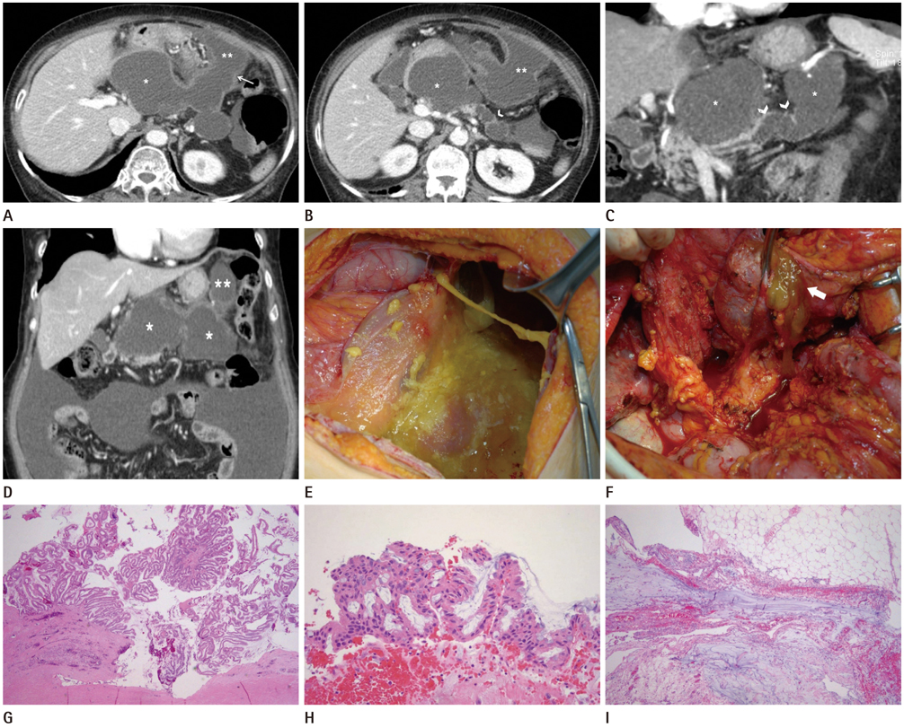
Pseudomyxoma Peritonei Caused by Ruptured Intraductal Papillary Mucinous Neoplasm of the Pancreas: A Case Report
- Affiliations
-
- 1Department of Radiology, Soonchunhyang University Bucheon Hospital, Soonchunhyang University College of Medicine, Bucheon, Korea. hklee@schmc.ac.kr
- 2Department of Pathology, Soonchunhyang University Bucheon Hospital, Soonchunhyang University College of Medicine, Bucheon, Korea.
- 3Department of Surgery, Soonchunhyang University Bucheon Hospital, Soonchunhyang University College of Medicine, Bucheon, Korea.
- KMID: 1941782
- DOI: http://doi.org/10.3348/jksr.2014.70.5.359
Abstract
- Pseudomyxoma peritonei (PMP) is an uncommon disease characterized by the seeding of mucin-secreting tumor cells throughout the abdomen and accumulation of mucin in the abdominal and pelvic cavities. Intraductal papillary mucinous neoplasms (IPMNs) of the pancreas are defined as pancreatic neoplasms that accumulate mucin within dilated ducts. Only a few cases of pancreatic IPMNs are associated with extra-pancreatic mucin and lead to PMP. This manuscript describes an unusual case of PMP caused by ruptured pancreatic IPMN.
Figure
Reference
-
1. van Ruth S, Acherman YI, van de Vijver MJ, Hart AA, Verwaal VJ, Zoetmulder FA. Pseudomyxoma peritonei: a review of 62 cases. Eur J Surg Oncol. 2003; 29:682–688.2. Ohashi K, Murakami Y, Maruyama M, Takekoshi T, Ohta H, Ohashi I. Four cases of mucous secreting pancreatic cancer. Prog Dig Endosc. 1982; 20:348–351.3. Zanelli M, Casadei R, Santini D, Gallo C, Verdirame F, La Donna M, et al. Pseudomyxoma peritonei associated with intraductal papillary-mucinous neoplasm of the pancreas. Pancreas. 1998; 17:100–102.4. Imaoka H, Yamao K, Salem AA, Mizuno N, Takahashi K, Sawaki A, et al. Pseudomyxoma peritonei caused by acute pancreatitis in intraductal papillary mucinous carcinoma of the pancreas. Pancreas. 2006; 32:223–224.5. Mizuta Y, Akazawa Y, Shiozawa K, Ohara H, Ohba K, Ohnita K, et al. Pseudomyxoma peritonei accompanied by intraductal papillary mucinous neoplasm of the pancreas. Pancreatology. 2005; 5:470–474.6. Nepka Ch, Potamianos S, Karadana M, Barbanis S, Kapsoritakis A, Koukoulis G. Ascitic fluid cytology in a rare case of pseudomyxoma peritonei originating from intraductal papillary mucinous neoplasm of the pancreas. Cytopathology. 2009; 20:271–273.7. Rosenberger LH, Stein LH, Witkiewicz AK, Kennedy EP, Yeo CJ. Intraductal papillary mucinous neoplasm (IPMN) with extra-pancreatic mucin: a case series and review of the literature. J Gastrointest Surg. 2012; 16:762–770.8. Brugge WR, Lauwers GY, Sahani D, Fernandez-del Castillo C, Warshaw AL. Cystic neoplasms of the pancreas. N Engl J Med. 2004; 351:1218–1226.9. Hinson FL, Ambrose NS. Pseudomyxoma peritonei. Br J Surg. 1998; 85:1332–1339.10. Tanaka M, Chari S, Adsay V, Fernandez-del Castillo C, Falconi M, Shimizu M, et al. International consensus guidelines for management of intraductal papillary mucinous neoplasms and mucinous cystic neoplasms of the pancreas. Pancreatology. 2006; 6:17–32.


