Radiologic Findings of the Pancreatic Lymphoepithelioma-Like Carcinoma: A Case Report
- Affiliations
-
- 1Department of Diagnostic Radiology, College of Medicine, Yeungnam University, Daegu, Korea. sungho1999@ynu.ac.kr
- 2Department of Pathology, College of Medicine, Yeungnam University, Daegu, Korea.
- KMID: 1941773
- DOI: http://doi.org/10.3348/jksr.2014.70.6.439
Abstract
- A 76-year-old man with fatigue was admitted to our hospital. Computed tomography (CT) and magnetic resonance imaging (MRI) showed a well-circumscribed mass with delayed heterogeneous enhancement between the pancreas head and the second portion of the duodenum. A surgical resection was performed and a lymphoepithelioma-like carcinoma (LEC) was pathologically confirmed, originating from the head of the pancreas. After the surgery, the patient did not have additional treatment and his LEC did not recur for four years and 10 months. We report MRI and CT findings with histopathologic findings of an extremely rare case of LEC arising from the pancreatic head.
Figure
-
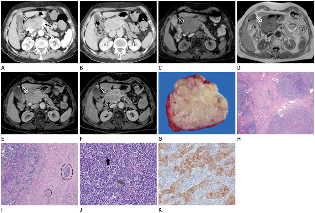
Fig. 1 Lymphoepithelioma-like carcinoma of the pancreas head in a 76-year-old man. A. Arterial phase image of abdominal CT shows an oval, well-defined, relatively homogeneous enhancement of the mass (white arrow) displacing adjacent vessel (black arrows). B. In the delayed phase scan, a more heterogeneously enhanced mass (white arrow) is noted. C. Axial T1-weighted gradient-recalled echo MR image shows a well-defined mass (white arrow) with intermediate to low signal intensity relative to the distal pancreas. D. Axial T2-weighted turbo-spin echo MR image shows mass (white arrow) with high signal intensity relative to the distal pancreas. E. Axial arterial-phase image of gadolinium-enhanced T1-weighted fat-suppressed gradient-recalled echo MR image shows relatively homogeneous enhancement and smooth contour of the mass (white arrow). F. Axial five-minute delayed MR image reveals a more heterogeneous mass (white arrow) with bright rim- and septal enhanced portions (black arrows) and poorly enhanced nodular portions. G. Cut surface of the gross specimen shows a well-circumscribed, lobulated and gray-white mass. H. The nests (white arrows) consisted of tumor cells and lymphoid cells are surrounded by fibrous tissue (hematoxylin and eosin stain, × 40). I. Photomicrograph of histologic specimen of the tumor shows atrophied pancreatic islets (black circles) in the periphery (hematoxylin and eosin stain, × 40). J. Photomicrograph of tumor cells (black arrow) shows an arranged syncytial aggregates of lymphoplasma cells (gray arrow) (hematoxylin and eosin stain, × 200). K. Tumor cells are positive for cytokeratin 19 (immunohistochemical stain, × 200).
Reference
-
1. Ishida M, Mori T, Shiomi H, Naka S, Tsujikawa T, Andoh A, et al. Non-Epstein-Barr virus associated lymphoepithelioma-like carcinoma of the inferior common bile duct. World J Gastrointest Oncol. 2011; 3:111–115.2. Kekis PB, Murtin C, Künzli BM, Kappler A, Buchholz B, Büchler MW, et al. Epstein-Barr virus-associated lymphoepithelial carcinoma in the pancreas. Pancreas. 2004; 28:98–102.3. Burke AP, Yen TS, Shekitka KM, Sobin LH. Lymphoepithelial carcinoma of the stomach with Epstein-Barr virus demonstrated by polymerase chain reaction. Mod Pathol. 1990; 3:377–380.4. Delaney D, Chetty R. Lymphoepithelioma-like carcinoma of the colon. Int J Clin Exp Pathol. 2012; 5:105–109.5. Dinniwell R, Hanna WM, Mashhour M, Saad RS, Czarnota GJ. Lymphoepithelioma-like carcinoma of the breast: a diagnostic and therapeutic challenge. Curr Oncol. 2012; 19:e177–e183.6. Williamson SR, Zhang S, Lopez-Beltran A, Shah RB, Montironi R, Tan PH, et al. Lymphoepithelioma-like carcinoma of the urinary bladder: clinicopathologic, immunohistochemical, and molecular features. Am J Surg Pathol. 2011; 35:474–483.7. Val-Bernal JF, González-Márquez P, Ballestero R, Zubillaga S. Primary lymphoepithelioma-like carcinoma of the ureter. Ann Diagn Pathol. 2011; 15:218–220.8. Kurihara K, Nagai H, Kasahara K, Kawai T, Saito K, Kanazawa K. Pleomorphic carcinoma of the pancreas with massive lymphocytic stromal infiltration and long-term survival after resection. Int J Pancreatol. 2000; 27:241–248.9. Wilentz RE, Goggins M, Redston M, Marcus VA, Adsay NV, Sohn TA, et al. Genetic, immunohistochemical, and clinical features of medullary carcinoma of the pancreas: a newly described and characterized entity. Am J Pathol. 2000; 156:1641–1651.
- Full Text Links
-
- Actions
-
Cited
- CITED
-
- Close
- Share
-
- Similar articles
-
- Lymphoepithelioma-like Carcinoma of the Renal Pelvis
- A Case of Lymphoepithelioma - Like Carcinoma of Uterine Cervix
- A Case of Lymphoepithelioma-Like Carcinoma in the Thyroid Gland
- Mixed lymphoepithelioma-like carcinoma and adenocarcinoma of the gallbladder
- A case of submucosal gastric lymphoepithelioma-like carcinoma
