J Korean Soc Radiol.
2014 Jun;70(6):408-410. 10.3348/jksr.2014.70.6.408.
Unusual Anatomical Variant of the Left Posterior Basal Segmental Pulmonary Artery: CT Findings
- Affiliations
-
- 1Department of Radiology, Sanggye Paik Hospital, Inje University College of Medicine, Seoul, Korea. s2621@paik.ac.kr
- 2Department of Emergency Medicine, Sanggye Paik Hospital, Inje University College of Medicine, Seoul, Korea.
- KMID: 1941766
- DOI: http://doi.org/10.3348/jksr.2014.70.6.408
Abstract
- To avoid vessel injury during minimally invasive surgery of the lung, exact knowledge of the pulmonary vasculature is important to surgeon. In this report, we present the case of unusual anatomical variant of the left posterior basal segmental pulmonary artery, arising from the left main pulmonary artery. This anomaly is rare but easily overlooked during interpretation of CT scans, potentially resulting in serious vessel injury during minimally invasive surgery.
Figure
-
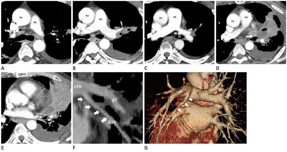
Fig. 1 A 52-year-old man with an incidentally detected unusual anatomical variant of the left posterior basal segmental pulmonary artery. A-E. Serial contrast enhanced axial chest CT scans show an anomalous vessel (arrows), directly arise from the posterior aspect of the left main pulmonary artery, anterior to the left main bronchus. F, G. Coronal maximum intensity projection image (F) and posterior oblique coronal volume rendered images (G) show an artery supplying left posterior basal segment (arrows), directly arise from left main pulmonary artery before branching upper lobar artery. Note.-Ao = ascending aorta, BT = truncus basalis, LB = left main bronchus, LPA = left pulmonary artery, PA = pulmonary artery
Reference
-
1. Yildirim A, Karabulut N, Dogğan S, Herek D. Congenital thoracic arterial anomalies in adults: a CT overview. Diagn Interv Radiol. 2011; 17:352–362.2. Yamada S, Inoue Y, Suga A, Iwazaki M. Surgical risk of vessel injury: an unusual anatomical variant of the right medial basal segmental pulmonary artery. Gen Thorac Cardiovasc Surg. 2011; 59:301–303.3. Goo HW, Park IS, Ko JK, Kim YH, Seo DM, Yun TJ, et al. CT of congenital heart disease: normal anatomy and typical pathologic conditions. Radiographics. 2003; 23 Spec No:S147–S165.4. Castañer E, Gallardo X, Rimola J, Pallardó Y, Mata JM, Perendreu J, et al. Congenital and acquired pulmonary artery anomalies in the adult: radiologic overview. Radiographics. 2006; 26:349–371.5. Le Brigand H. Nouveau traite de technique chirurgicale (tome III). Paris: Masson et Cie;1973. p. 304–305.6. Matsumoto K, Yamasaki N, Tsuchiya T, Miyazaki T, Tomoshige K, Hayashi H, et al. Three-dimensional computed tomography for a mediastinal basal pulmonary artery. Ann Thorac Surg. 2012; 94:e115–e116.7. Kataoka K, Nishikawa T, Fujiwara T, Matsuura M. A case of lung cancer with an extremely rare branching pattern of the left A5+8+9+10 pulmonary artery. Jpn J Lung Cancer. 2010; 50:362–365.8. Shibano T, Endo S, Tetsuka K, Kanai Y. Dangerous mediastinal basal pulmonary artery during left upper lobectomy. Interact Cardiovasc Thorac Surg. 2011; 13:358–360.9. Moriyama S, Miyoshi K, Tada A, Kurosaki T. A case report of abnormal branching of left A8+9 pulmonary artery. Jpn J Chest Surg. 2009; 23:58–61.10. Sano M, Mizuno T, Iizuka M, Yamada T, Kasugai T, Ishiguro H. [Abnormal branching of left pulmonary artery to the lateral and posterior basal segments]. Nihon Kyobu Geka Gakkai Zasshi. 1996; 44:1772–1775.
- Full Text Links
-
- Actions
-
Cited
- CITED
-
- Close
- Share
-
- Similar articles
-
- Aberrant Origin of the Upper Left Lobe Anterior and Superior Lingular Segmental Pulmonary Artery Arising from the Right Pulmonary Artery: A Case Report
- Intralobar pulmonary sequestration with aberrant systemic artery - pulmonary vein fistula
- Pulmonary Artery Sling with Situs Solitus Dextroposition of Heart and Left Superior Vena Cava
- Left Bronchial Artery Arising from a Replaced Left Hepatic Artery in a Patient with Massive Hemoptysis
- Unusual Presentation of Extralobar Pulmonary Sequestration: A Case Report
