J Korean Soc Spine Surg.
2005 Sep;12(3):233-237. 10.4184/jkss.2005.12.3.233.
A Solitary Osteochondroma of the Pediatric Thoracic Spine: A Case Report
- Affiliations
-
- 1Department of Orthopaedic Surgery, Bundang CHA Hospital, College of Medicine, Pochon CHA University, Sung-Nam, Korea. hs55hong@hanmail.net
- 2Department of Pathology, Bundang CHA Hospital, College of Medicine, Pochon CHA University, Sung-Nam, Korea.
- KMID: 1941690
- DOI: http://doi.org/10.4184/jkss.2005.12.3.233
Abstract
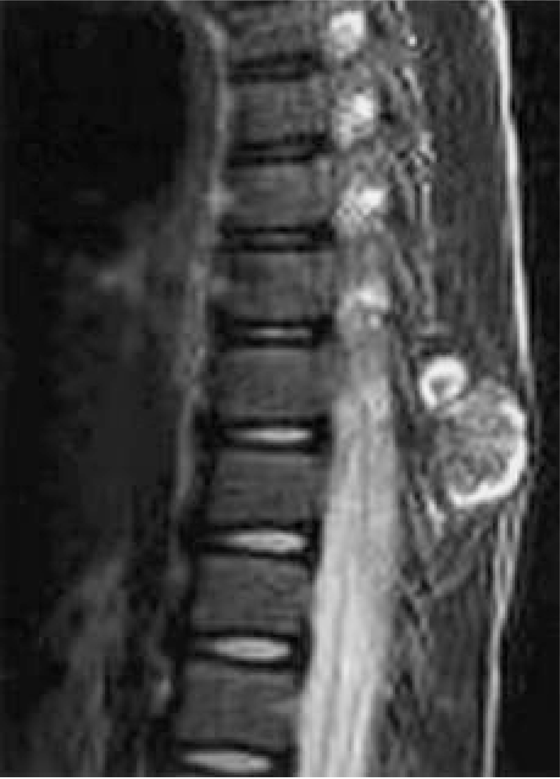
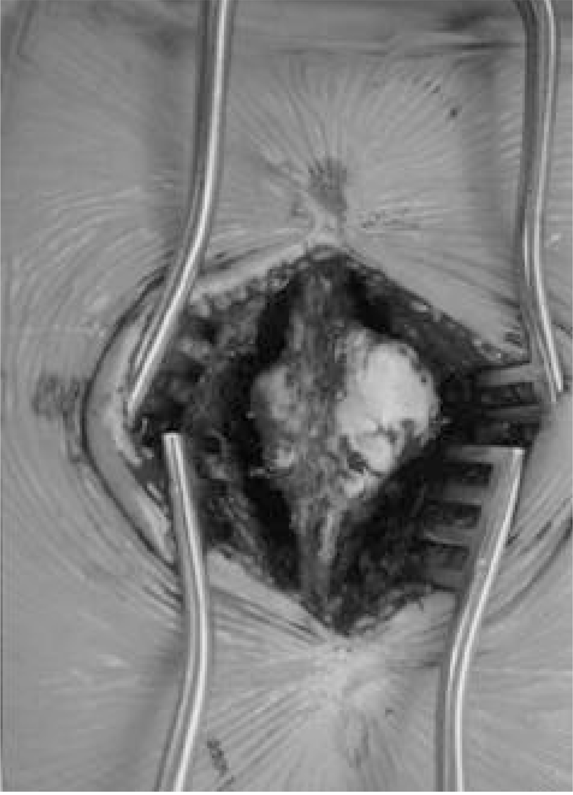
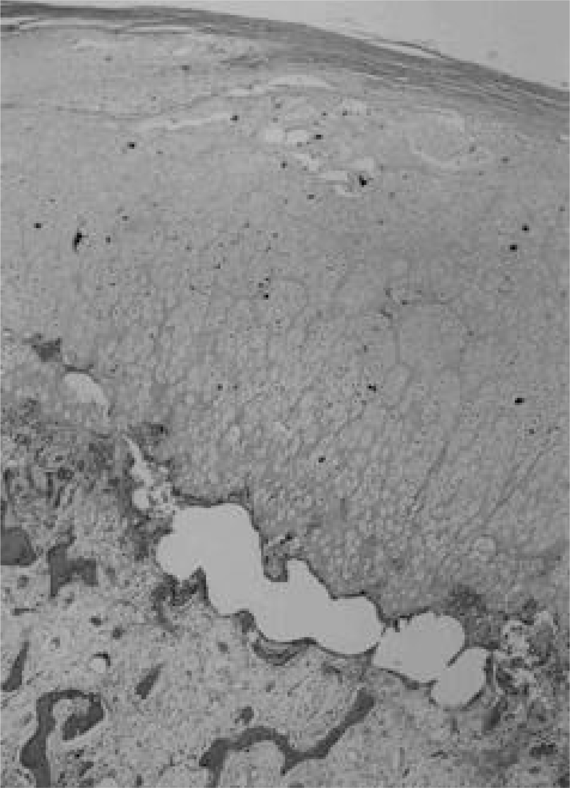
- An osteochondroma is a bone tumor, but rarely occurs in the thoracic spine, especially in the pediatric population. The objective of this study was to describe the diagnosis and successful treatment of a pediatric patient with an osteochondroma of the thoracic spinous process. The anteroposterior and lateral plain radiographs illustrated a well-defined solid mass arising from the spinous process of the tenth thoracic vertebrae. Computed tomography and magnetic resonance imaging further delineated that the mass arose from the spinous process, but with no obvious impingement of the nerve roots. After excision of the lesion, the gross pathological and histological evaluations were consistent with those of an osteochondroma. This led to appropriate surgical intervention, resulting in definitive treatment.
Keyword
Figure
Reference
-
1). Albrecht S, Crutchfield JS, SeGall GK. On spinal osteochondromas. J Neurosurg. 1992; 77:247–252.
Article2). Dahlin DC, Unni KK, In. Bone Tumors: General Aspects and Data on 11, 087 cases. 5th ed.Springfield, IL: Lippincott-Raven;1996. 11-25.3). Arasil E, Ederm A, Yuceer N. Osteochondroma of the upper cervical spine: a case report. Spine. 21:1996; 516–518.4). Cohen DM, Dahlin DC, Mac Carty CS. Apparently soli - tary tumors of the vertebral column. Mayo Clin Proc. 39:1964; 509–528.5). Elsberg CA. Surgical Diseases of the Spinal Cord, Membrane, and Nerve Roots: Symptoms, Diagnosis, and Treatment. New York: PB Hoeber. 1941; 359-360.6). Schmale GA, Conrad EU, Raskind WH. The natural history of hereditary multiple exostoses. J Bone J Surg. 76A:1994; 986–992.
Article7). Cooke SR, Cumming WJK, Cowie RA. Osteochondroma of the cervical spine: case report and review of the literature. Br J Neurosurg. 8:1994; 359–363.
Article8). Di Lorenzo N, Nardi P, Ciappetta P, Fortuna A. Benign tumors and tumorlike conditions of the spine. Surg Neurol. 25:1986; 449–456.9). Bullough PG. Orthopedic Pathology. 3rd ed.London;p. Mosby–Wolfe. 354-356:1997.10). Kenny PJ, Gilula LA, Murphy WA. The use of computed tomography to distinguish osteochondroma from chon -drosarcoma. Radiology. 139:1981; 129–137.11). Malat I, Virapongse C, Levine A. Solitary osteochondroma of the spine. Spine. 11:1986; 625–628.
Article12). Mirra JM, Picci P, Gold RH. Bone Tumors: Clinical, Radiologic, and Pathologic Correlations. Philadelphia: Lea & Febiger. 1989; 1626-1629.13). Linkowski GD, Tsai FY, Recher L, et al. Solitary osteochondroma with spinal cord compression. Surg Neurol. 23:1985; 388–390.
Article14). Chiurco AA. Multiple exostoses of bone with fatal spinal cord compression. Neurology. 20:1970; 275–278.
- Full Text Links
- Actions
-
Cited
- CITED
-
- Close
- Share
- Similar articles
-
- Solitary Osteochondroma of the Thoracic Spine Presenting as Spinal Cord Compression: Case Report
- Osteochondroma of 12th Thoracic Vertebra: A Case Report
- Osteochondroma of the Cervical Spine: A Case Report
- Osteochondroma of the Sacrum: A Case Report
- Solitary Spinal Osteochondroma Presenting as a Neck Mass: Case Report


