J Korean Soc Spine Surg.
2007 Jun;14(2):115-119. 10.4184/jkss.2007.14.2.115.
Congenital Absence of a Pedicle of L4 in the Spinal Stenosis: A Case Report
- Affiliations
-
- 1Department of Orthopedic Surgery, Spine Center Soonchunhyang University College of Medicine, Seoul, Korea. schsbj@hosp.sch.ac.kr
- KMID: 1941665
- DOI: http://doi.org/10.4184/jkss.2007.14.2.115
Abstract
- Congenital absence of a lumbar pedicle is an uncommon anomaly, and most cases are asymptomatic and discovered incidentally. A 72-year-old man presented with lower back pain that radiated to his bilateral lower extremities. Physical examination revealed no neurological deficits. Plain radiographs of the lumbar spine revealed absence of the left L4 pedicle, along with hypertrophy and sclerosis of the contralateral pedicle. Magnetic resonance imaging showed stenosis of the L3-4 neural canal. Computed tomography revealed absence of the left L4 pedicle associated with hypertrophy and sclerosis of the right L4 pedicle and facet joint. The symptoms of the patient were resolved after posterior decompression without fusion. Here, we report one case of congenital absence of an L4 pedicle detected in a spinal stenosis patient who need to undergo a decompressive surgery for the spinal stenosis caused by contralateral facet hypertrophy.
MeSH Terms
Figure
-
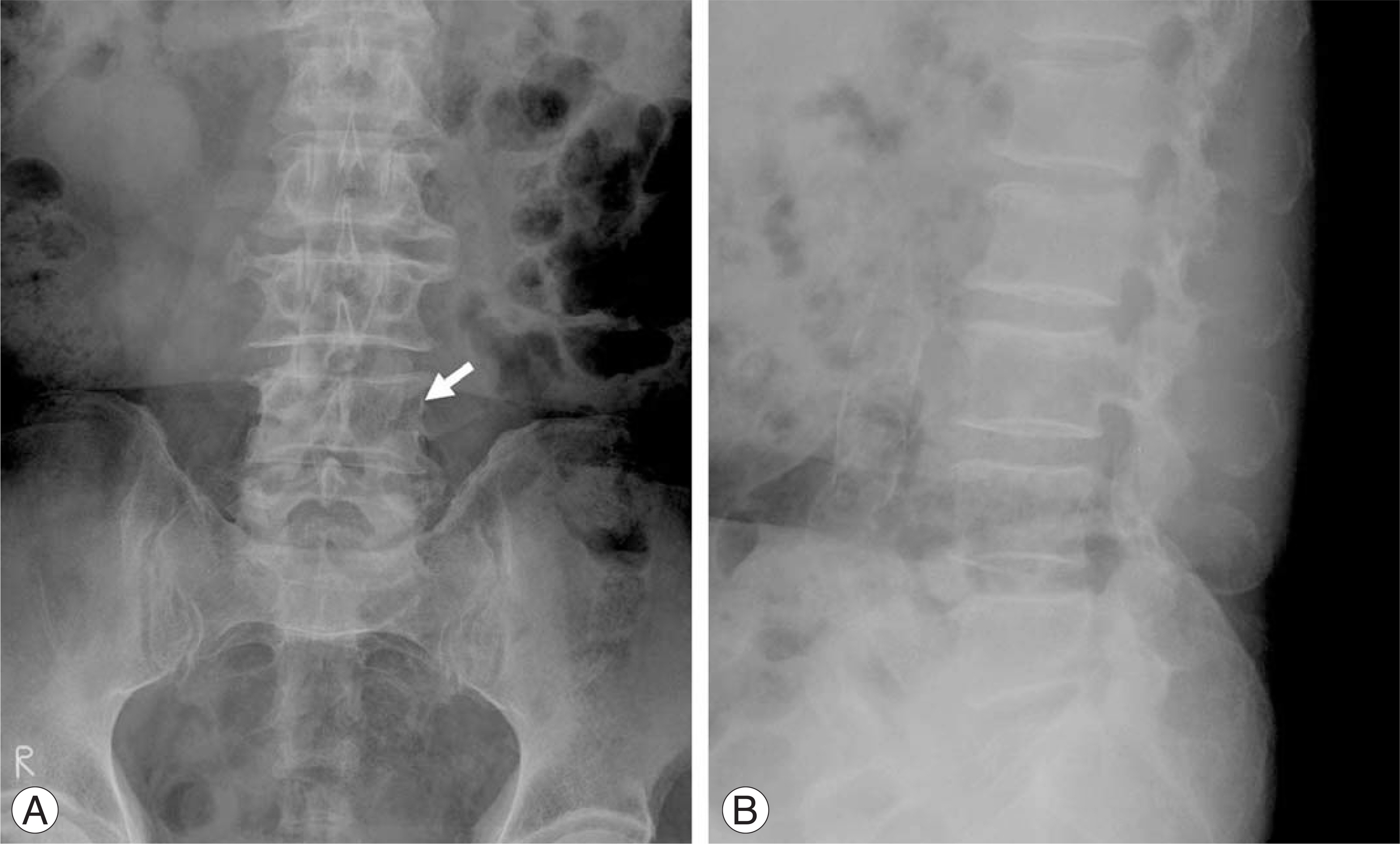
Fig. 1. Preoperative AP (A) and lateral (B) radiographs of a 72-year-old man suffering from spinal stenosis at L3-4 level. Arrow indicates absence of the left L4 pedicle.
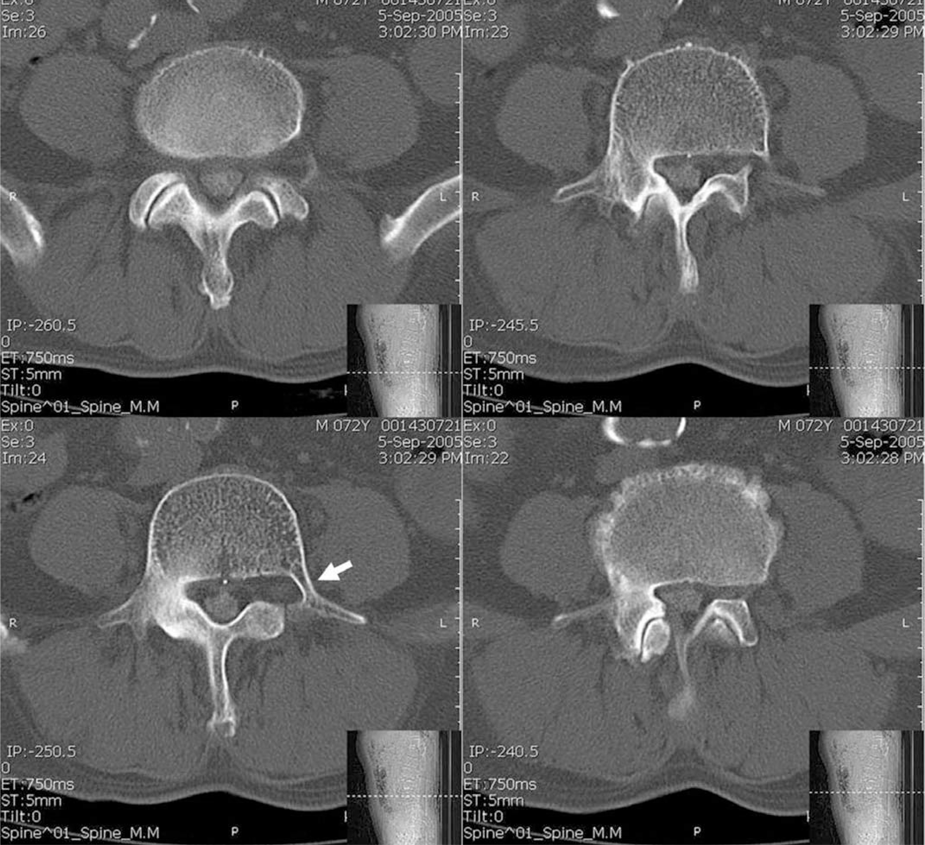
Fig. 2. CT scans shows absence of the left pedicle and hypertrophy of the right pedicle of L4 vertebra. Degenerative changes of the right L4-5 facet joint was also noted. Arrow indicates the rudimentary bony connection between the vertebral body and the left side transverse process, instead of the pedicle.
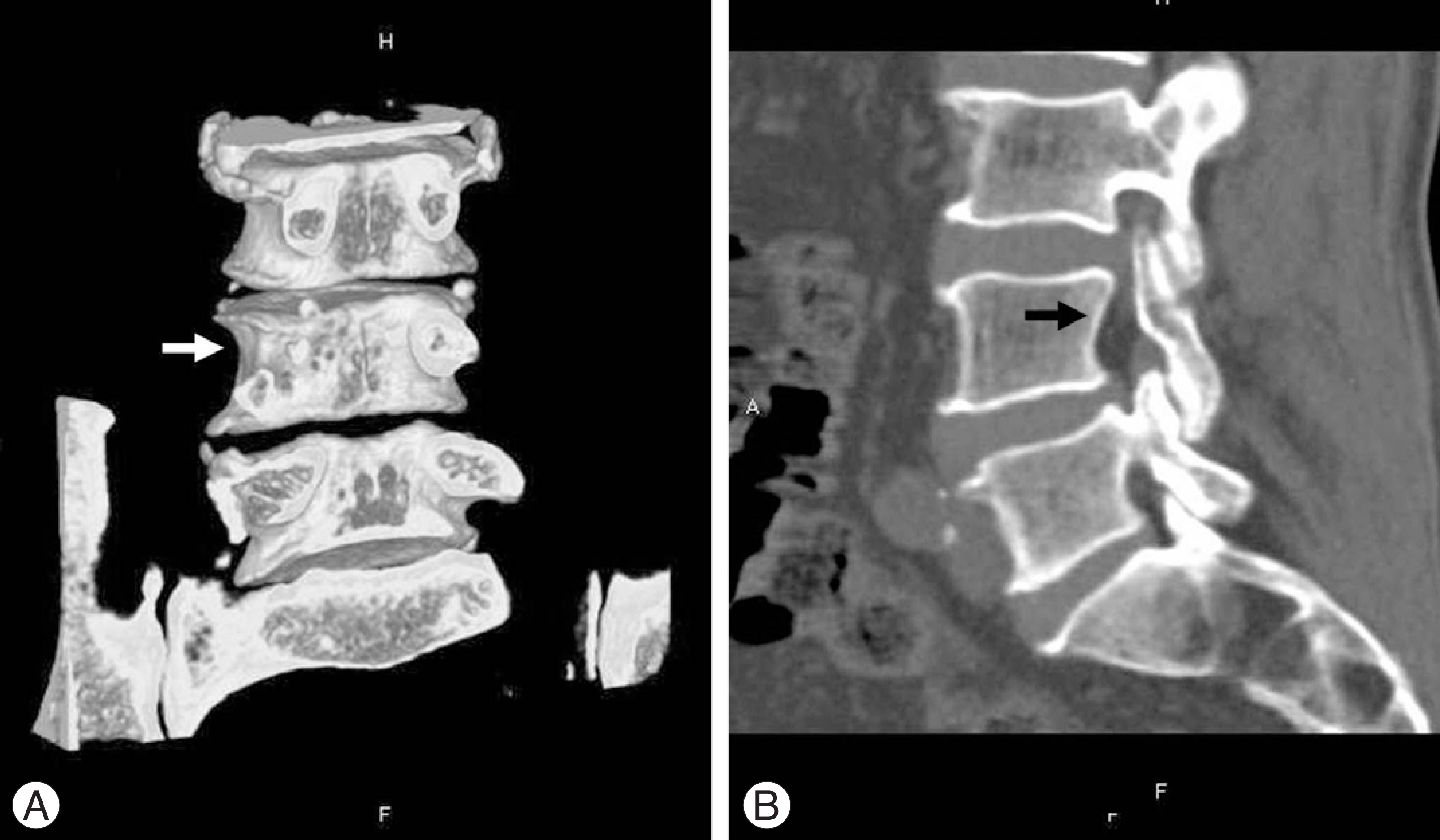
Fig. 3. Three-dimensional (A) & 2-D (B) reconstructed CT image shows absence of the left pedicle at L4 (arrow-pointed).
Reference
-
1). Kaito T, Kato Y, Sakaura H, Yamamoto K, Hosono N. Congenital absence of a lumbar pedicle presenting with contralateral lumbar radiculopathy. J Spinal Disord Tech. 2005; 18:203–205.
Article2). Mizutani M, Yamamuro T, Shikata J. Congenital absence of a lumbar pedicle. Spine. 1989; 14:890–891.
Article3). Villas C, Barrios RH. Congenital absence of the pedicles and the neural arch of L2. Eur Spine J. 1997; 6:354–356.
Article4). Lorenz M, Patwardhan A, Vanderby R. Load-bearing characteristics of lumbar facets in normal and surgically altered spinal segments. Spine. 1983; 8:122–130.
Article5). Sharma M, Langrana NA, Rodriguez J. Role of ligaments and facets in lumbar spinal stability. Spine. 1995; 20:887–900.
Article6). Abumi K, Panjabi MM, Kramer KM, et al. .:. Biomechanical evaluation of lumbar spinal stability after graded facetectomies. Spine. 1990; 15:1142–1147.
Article7). Kornberg M. Spondylolithesis with unilateral pars inter-articularis defect and contralateral facet joint degeneration. Spine. 1988; 13:712–713.8). Park KW, Kim BH, Lee JH, Song KS, Lee CK, Chang BS. Congenital absense of thoracic spine pedicle: Case report. J Korean Soc Spine Surg. 2006; 13:219–223.
- Full Text Links
-
- Actions
-
Cited
- CITED
-
- Close
- Share
-
- Similar articles
-
- Congenital Absence of Thoracic Spine Pedicle: Case Report
- Congenital Absence of a Cervical Spine Pedicle : Report of Two Cases and Review of the Literature
- Fusion of Pedicular Cleft Using Pedicle Screw Fixation: A Case Report
- Intradural Schwannoma Associated with Lumbar Spinal Stenosis: A Case Report
- Cotrel - Dubousset Pedicle Screw Fixation After Posterior Decompression of Lumbar Spinal Stenosis
