Korean J Leg Med.
2014 Nov;38(4):171-174. 10.7580/kjlm.2014.38.4.171.
Diver Death due to Underwater Explosion
- Affiliations
-
- 1Forensic Medicine Division, Gwangju Institute, National Forensic Service, Jangseong, Korea. pdrdream@gmail.com
- 2Physical Engineering Division, Gwangju Institute, National Forensic Service, Jangseong, Korea.
- 3Forensic Chemistry Division, Gwangju Institute, National Forensic Service, Jangseong, Korea.
- 4Medical Examiner's Office, National Forensic Service, Wonju, Korea.
- 5Department of Forensic Medicine, Chosun University School of Medicine, Gwangju, Korea.
- KMID: 1928011
- DOI: http://doi.org/10.7580/kjlm.2014.38.4.171
Abstract
- A 44-year-old man was cutting an outer plate of a ship, at a depth of 25 m below sea level. Following a sudden explosion, he was discovered unconscious and was carried to the surface by other divers. There was no evidence of vital signs upon arrival at the hospital. Postmortem computed tomography, which was performed prior to autopsy, revealed massive pneumocephalus in the brain, pneumohemothorax, diffuse lung contusions with multiple traumatic lung cysts, air-fluid level in the cardiac chamber of the chest, and pneumoperitoneum in the abdomen. Postmortem external examination showed a circular abrasion on the jaw, diffuse subcutaneous emphysema, and contusion in the right upper arm. An internal examination revealed intravascular air bubbles in all four chambers of the heart, and diffuse pulmonary trauma including contusion, laceration, and multiple traumatic cysts. Blast injury to the chest, and air embolism due to the underwater explosion were established as the underlying cause of death.
Keyword
MeSH Terms
Figure
-
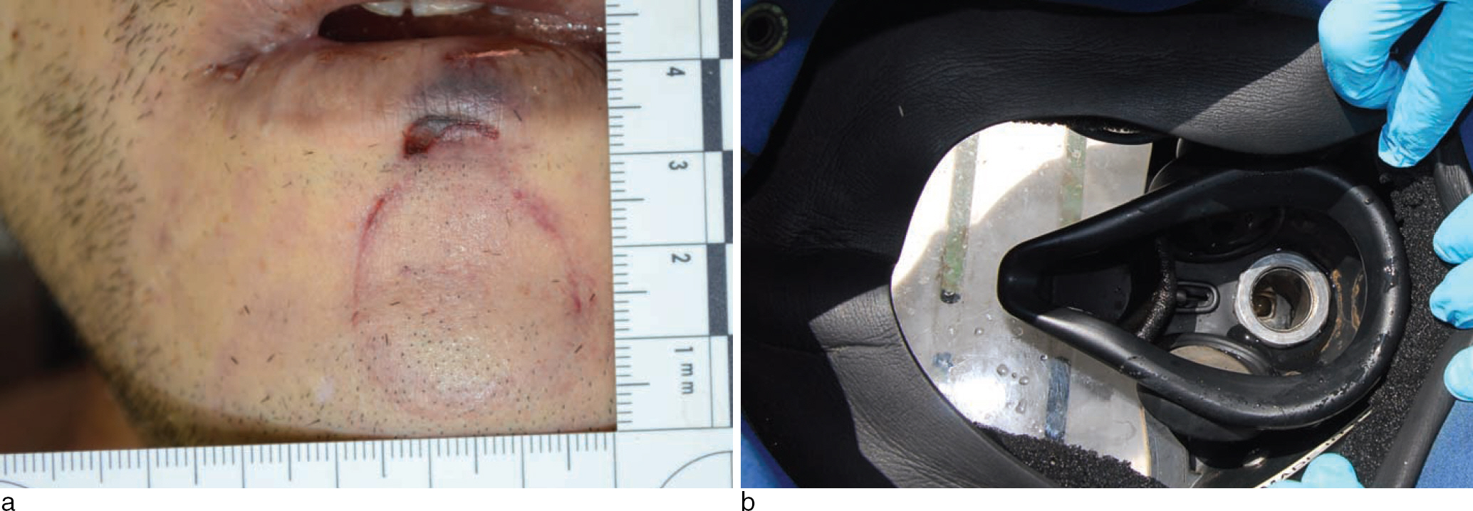
Fig. 1. Brain computed tomography shows a massive pneumocephalus and air in the cerebral vessels and subcutaneous tissue (a). Chest computed tomography shows a massive subcutaneous emphysema, pneumo-hemothorax, pneumomediastinum, multiple traumatic lung cysts, and air fluid levels in cardiac chamber and aorta (b).
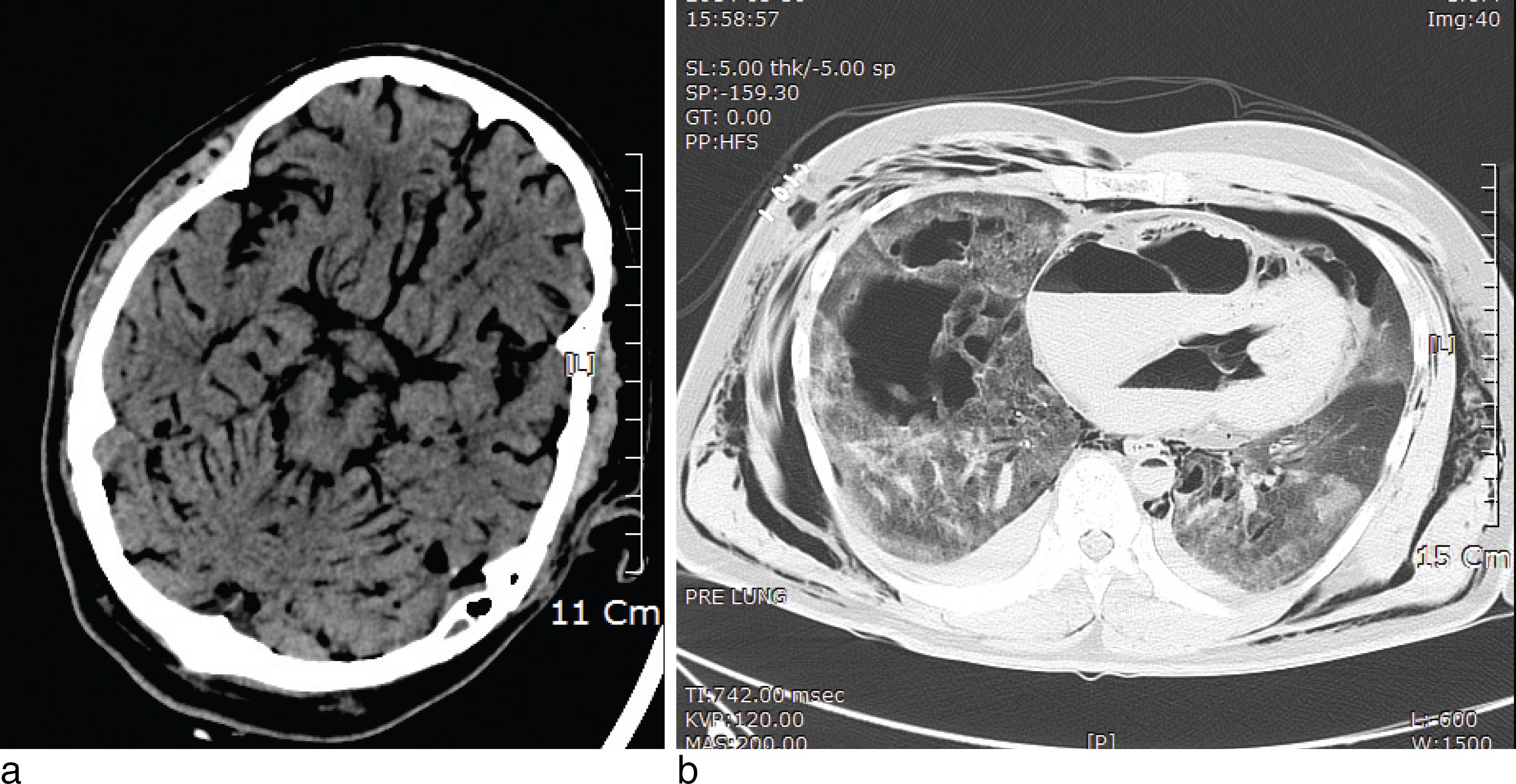
Fig. 2. A circular abrasion sized about 2.5 cm in diameter is noted in his jaw (a). And a similar sized circular air-orifice is noted in the diving helmet (b).
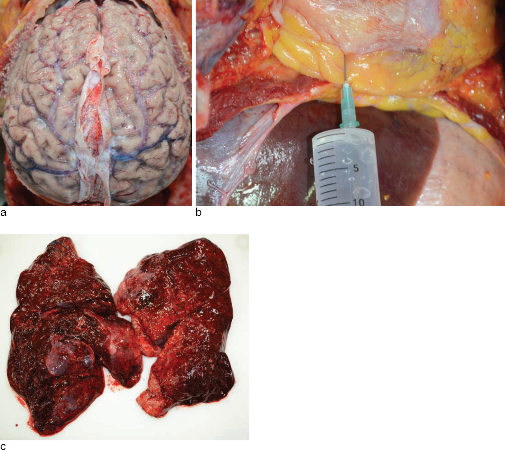
Fig. 3. Gas bubbles present in the superficial cerebral blood vessels (a). Gas bubbles are seen in the heart chambers (b). A massive pulmonary traumas including laceration, hilar rupture, multiple hemorrhagic cysts, and contusion are noted (c).
Reference
-
References
1. Kim YS. Technical approach for the postmortem examination of SCUBA diving fatality. Korean J Leg Med. 2014; 38:1–7.
Article2. Ihama Y, Miyazaki T, Fuke C, et al. Scuba-diving related deaths in Okinawa, Japan, from 1982 to 2007. Legal Med (Tokyo). 2008; 10:119–24.
Article3. Saukko P, Knight B. Knight's forensic pathology. 3rd ed.London: Arnold;2004. p. 274–5.4. Turkmen N, Akan O, Cetin S, et al. Scuba diver deaths due to air embolism: two case reports. Soud Lek. 2013; 58:26–8.5. Lawrence C. Interpretation of gas in diving autopsies. SPUMS J. 1997; 27:228–30.6. Wheen LC, Williams MP. Postmortems in recreational s-cuba diver deaths: the utility of radiology. J Forensic Leg Med. 2009; 16:273–6.
Article
- Full Text Links
-
- Actions
-
Cited
- CITED
-
- Close
- Share
-
- Similar articles
-
- Pulmonary Involvement in Decompression Sickness of a Self Contained Underwater Breath Apparatus Diver
- SCUBA Diving Fatality by Overweighted Belt: An Autopsy Case
- Forensic Review of Underwater Diving-Related Death
- A Primer to Diving Physiology
- Checklist for Forensic Investigation on Fatal SCUBA Diving Accidents