Prog Med Phys.
2013 Sep;24(3):176-182. 10.14316/pmp.2013.24.3.176.
Enhancement of the Early/Precise Diagnosis Based on the Measurement of SUVs in F-18 FDG PET/CT Whole-body Image
- Affiliations
-
- 1Department of Radiologic Technology, Daegu Health College, Daegu, Korea.
- 2Department of Radiation Oncology, Yeungnam University College of Medicine, Daegu, Korea. skkim3@ynu.ac.kr
- 3Department of Nuclear Medicine, Yeungnam University College of Medicine, Daegu, Korea.
- 4Department of Radiology, Soonchunhyang University of Hospital Gumi, Gumi, Korea.
- KMID: 1910582
- DOI: http://doi.org/10.14316/pmp.2013.24.3.176
Abstract
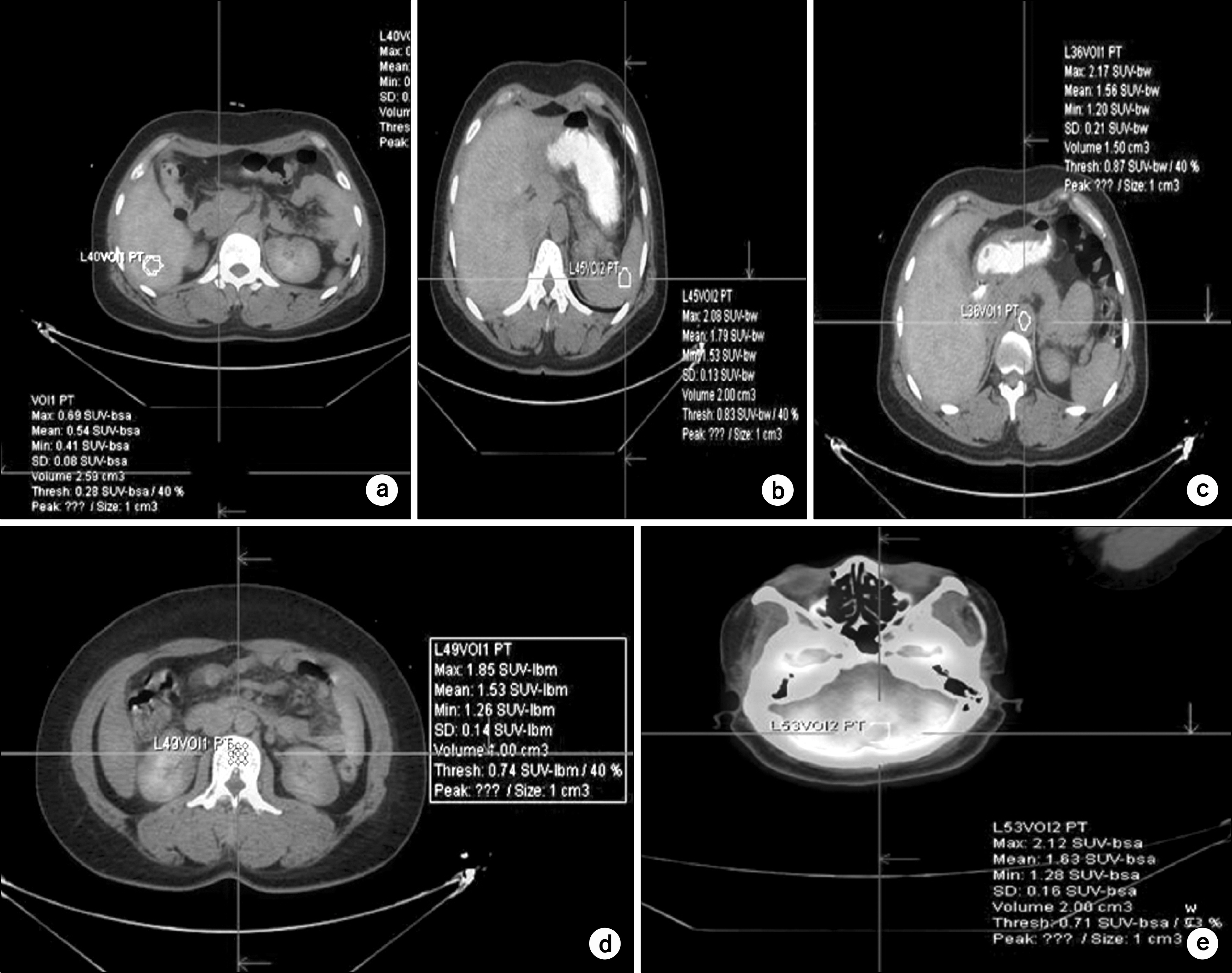
- Through this research, we measure the data for several SUVs such as SUVLBM, SUVBW, and SUVBSA using volume of interest in order to enhance the diagnostic level in whole-body image for healthy examinees via F-18 FDG PET/CT. Maximum value, mean value, standard deviation, and threshold value for each SUVs are shown. The measurement of SUVs are carried out with 31 examinees who have taken whole-body examination with F-18 FDG PET/CT from July, 2012 to August, 2012. To secure the preciseness of measurement, we selected 26 healthy examinees as a subject of measurement according to diagnostic view of a nuclear-medical doctor. We see from the measurement of SUVs of PET/CT that the value of SUVBW is hightest and followed by SUVLBM and SUVBSA in turn regardless of the use of contrast media. By comparing the SUVLBM-maximum data for the group used contrast media with those for the group used no contrast media, there found a trend that the measured values increase when the contrast media are used. Among them, liver, aorta, lumbar-5, and Cerebellum exhibit significant difference (p<0.05). We conclude that our data for SUVs would be basic references in overall image interpretation, and hope that the research using VOI would be active.
Keyword
Figure
Cited by 1 articles
-
Measurement and Estimation for the Clearance of Radioactive Waste Contaminated with Radioisotopes for Medical Application
Changbum Kim, MinSeok Park, Gi-sub Kim, Haijo Jung, Seongjoo Jang
Prog Med Phys. 2014;25(1):8-14. doi: 10.14316/pmp.2014.25.1.8.
Reference
-
1. Park SY. Consideration on the satisfaction of patients and Variation according to whether or not to listen to music after F-18 FDG Injection. The Graduate School of Bio-Medical Science, Korea University. 2013.2. Oriuchi N, Higuchi T, Ishikita T, et al. Present role and future prospects of positron emission tomography in clinical oncology. Cancer Sci. 97(12):1291–1297. 2006.
Article3. Hany TF, Steinert HC, Thomas F. Integrated PET/CT: current applications and future direction. Radiology. 238::. (1):405–422. 2006.4. Benamor M, Ollivier L, Brisse H, Moulin R, Servois V, Neuenschwander S. The clinical role of CT/PET in oncology: an update. Cancer Imaging. 5::. (8):68–75. 2005.
Article5. Bar-Shalom R, Yefremov N, Guralnik L, et al. Clinical performance of PET/CT in evaluation of cancer: additional value for diagnostic imaging and patient management. The Journal of Nuclear Medicine. 44::. (9):1200–1209. 2013.6. Weber WA. Positron emission tomography as an imaging biomaker. J Clin Oncol. 24(20):3282–3292. 2006.7. Czernin J, Allen M, Schelbert R. Improvements in cancer staging with PET/CT: Literature based evidence as of September. J Nucl Med. 48(1):78–88. 2007.8. Tian M, Zhang H, Nakasone Y, Mogi K, Endo K. Expression of Glut-1 and Glut-3 in untreaed oral squamous cell carcinoma compared with FDG accumulation in a PET study. 31(1):5–12. 2004.9. Tohma T, Okazumi S, Makino H, et al. Relationship between glucose transporter, hexokinase and FDG-PET in esophageal cancer. Hepato-Gastroenterol. 52::. (62):486–490. 2005.10. Zincirkeser S, Şahin E, Halac M, Sageret S. Standardized uptake values of normal organs on 18F-Fluorodeoxyglucose positron emission tomography and computed tomography imaging. J Int Med Res. 35::. (2):231–236. 2007.
Article11. Boellaard R. Standards for PET image acquisition and quantitative data analysis. J Nucl Med. 50(1):11–20. 2009.
Article12. Lee HS. A study for distortion of standardized uptake value according to the does and lesion size using 18F-FDG PET/CT. Graduates School Korea Univ. 2012.13. Bushnell D, Madsen M, Menda Y, et al. Evaluation of various corrections to the standardized uptake value for diagnosis of pulmonary malignancy. Nucl Med Common. 22(1):1077–1081. 2001.14. Zasadny KR, Wahl RL. Standardized uptake values of normal tissues at PET with2-[fluorine-18]-fluoro-2-deoxy-D-glucose: variations with body weight and a method for correction. Radiology. 189::. (1):847–850. 1993.15. Wahl RL, Jacene H, Kasamon Y, Lodge MA. From RECIST to PERCIST: evolving considerations for PET response criteria in solid tumors. JNM. 50::. (1):122–149. 2009.
Article16. Eiber M, Martinez-Möller A, Souvatzoglou M, et al. Value of a Dixon-based MRI/PET attenuation correction sequence for the localization and evaluation of PET-positive lesions. Eur J Nucl Med Mol Imaging. 38::. (5):1691–1701. 2011.17. Boellaard R, O'Doherty Mike J, Weber Wolfgang A, et al. FDG PET and PET/CT: EANM procedure guidelines for tumour PET imaging: version 1.0. Eur J Nucl Med Mol Imaging. 37::. (10):181–200. 2010.
- Full Text Links
- Actions
-
Cited
- CITED
-
- Close
- Share
- Similar articles
-
- The Proper Use of PET/CT in Tumoring Imaging
- Usefulness of 18 F-FDG PET/CT and Multiphase CT in the Differential Diagnosis of Hepatocellular Carcinoma and Combined Hepatocellular CarcinomaCholangiocarcinoma
- Whole Body Positron Emission Tomography/Computed Tomography
- A Case of Incidentally Detected Nasopharyngeal Tuberculosis on F-18 FDG PET/CT
- Primary Hepatosplenic B-cell Lymphoma: Iinitial Diagnosis and Assessment of Therapeutic Response with F-18 FDG PET/CT


