J Korean Soc Surg Hand.
2014 Dec;19(4):180-188. 10.12790/jkssh.2014.19.4.180.
The Results of Conservative Management for Early Stage Kienbock's Disease
- Affiliations
-
- 1Department of Orthopaedic Surgery, Samsung Medical Center, Sungkyunkwan University School of Medicine, Seoul, Korea. mjp3506@skku.edu
- KMID: 1896907
- DOI: http://doi.org/10.12790/jkssh.2014.19.4.180
Abstract
- PURPOSE
Early stage Kienbock's disease is commonly treated with a surgical intervention to avoid progression to degenerative change. The purpose of this study was to evaluate the clinical and radiographic outcomes of conservative management in patients with an early stage Kienbock's disease with a hypothesis that the lunate can be maintained in patients with no pain or tolerable pain.
METHODS
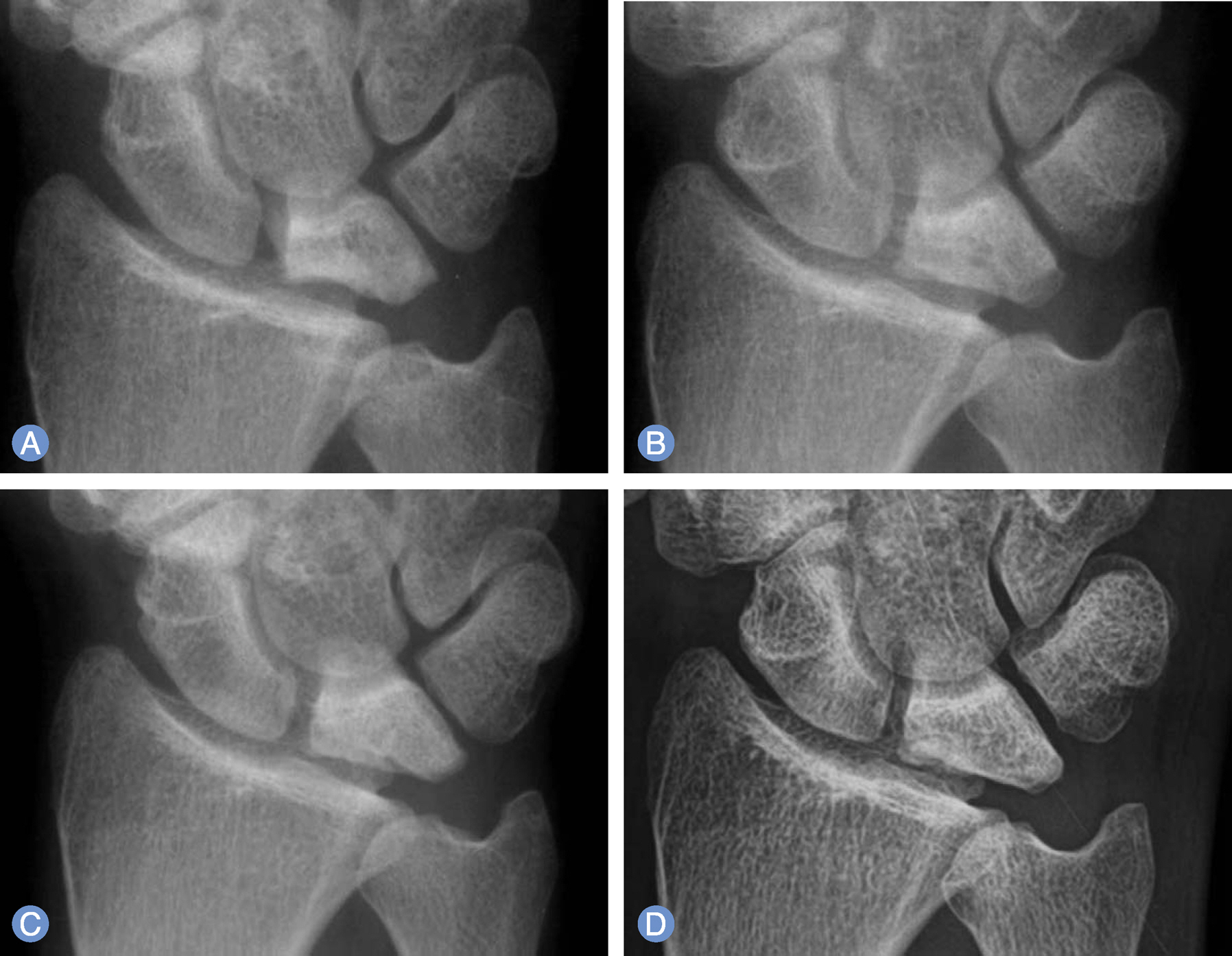
Twenty-three patients with a Lichtman stage I, II or IIIA Kienbock's disease were managed conservatively and investigated prospectively. There were ten men and thirteen women. Mean age at first visit was 53.9 years old and mean follow-up period was 51.3 months. The clinical outcomes were evaluated by range of motion, subjective satisfaction of patients at final follow-up. Radiographic measurements of the Lichtman stage were assessed at first visit and at the final follow-up. Three patients were Lichtman stage I, eleven patients were II and nine patients were IIIA.
RESULTS
Range of motion improved in all cases. According to Dornan's criteria, eleven patients were excellent, another eleven patients were good and one patient was fair. Based on Lichtman stage, no change was seen in sixteen patients, while seven showed progression. Three patients revealed improved radiographic findings of the lunate at final follow-up.
CONCLUSION
We found that conservative management including close observation of clinical and radiographic changes can provide satisfactory clinical improvement in patients with no pain or tolerable pain in early stage Kienbock's disease.
Keyword
MeSH Terms
Figure
Reference
-
1. Lichtman DM, Degnan GG. Staging and its use in the determination of treatment modalities for Kienbock's disease. Hand Clin. 1993; 9:409–16.2. Allan CH, Joshi A, Lichtman DM. Kienbock's disease: diagnosis and treatment. J Am Acad Orthop Surg. 2001; 9:128–36.3. Beredjiklian PK. Kienbock's disease. J Hand Surg Am. 2009; 34:167–75.4. Mirabello SC, Rosenthal DI, Smith RJ. Correlation of clinical and radiographic findings in Kienbock's disease. J Hand Surg Am. 1987; 12:1049–54.5. Delaere O, Dury M, Molderez A, Foucher G. Conservative versus operative treatment for Kienbock's disease. A retrospective study. J Hand Surg Br. 1998; 23:33–6.6. Kristensen SS, Thomassen E, Christensen F. Kienbock's disease: late results by non-surgical treatment. A follow-up study. J Hand Surg Br. 1986; 11:422–5.7. Dornan A. The result of treatment of Kienbock's disease. J Bone Joint Surg Br. 1949; 31:518–20.8. Lichtman DM, Mack GR, MacDonald RI, Gunther SF, Wilson JN. Kienbock's disease: the role of silicone replacement arthroplasty. J Bone Joint Surg Am. 1977; 59:899–908.9. Moran SL, Cooney WP, Berger RA, Bishop AT, Shin AY. The use of the 4+5 extensor compartmental vascularized bone graft for the treatment of Kienbock's disease. J Hand Surg Am. 2005; 30:50–8.10. Salmon J, Stanley JK, Trail IA. Kienbock's disease: conservative management versus radial shortening. J Bone Joint Surg Br. 2000; 82:820–3.11. Ozalp T, Yercan HS, Okcu G. The treatment of Kienbock disease with vascularized bone graft from dorsal radius. Arch Orthop Trauma Surg. 2009; 129:171–5.12. Innes L, Strauch RJ. Systematic review of the treatment of Kienbock's disease in its early and late stages. J Hand Surg Am. 2010; 35:713–717.e4.13. Mennen U, Sithebe H. The incidence of asymptomatic Kienbock's disease. J Hand Surg Eur Vol. 2009; 34:348–50.14. Lichtman DM, Lesley NE, Simmons SP. The classification and treatment of Kienbock's disease: the state of the art and a look at the future. J Hand Surg Eur Vol. 2010; 35:549–54.15. Taniguchi Y, Tamaki T, Honda T, Yoshida M. Rotatory subluxation of the scaphoid in Kienbock's disease is not a cause of scapholunate advanced collapse (SLAC) in the wrist. J Bone Joint Surg Br. 2002; 84:684–7.16. Luo J, Diao E. Kienbock's disease: an approach to treatment. Hand Clin. 2006; 22:465–73.17. Taniguchi Y, Nakao S, Tamaki T. Incidentally diagnosed Kienbock's disease. Clin Orthop Relat Res. 2002; (395):121–7.18. Dias JJ, Lunn P. Ten questions on Kienbock's disease of the lunate. J Hand Surg Eur Vol. 2010; 35:538–43.19. Schmitt R, Heinze A, Fellner F, Obletter N, Struhn R, Bautz W. Imaging and staging of avascular osteonecroses at the wrist and hand. Eur J Radiol. 1997; 25:92–103.
Article20. Arnaiz J, Piedra T, Cerezal L, et al. Imaging of Kienbock disease. AJR Am J Roentgenol. 2014; 203:131–9.


