Ann Surg Treat Res.
2014 May;86(5):270-273. 10.4174/astr.2014.86.5.270.
Chronic gastric anisakiasis provoking a bleeding gastric ulcer
- Affiliations
-
- 1Department of Surgery, Digestive Disease Research Institute and Institute of Medical Science, Wonkwang University College of Medicine, Iksan, Korea. rjk@wonkwang.ac.kr
- KMID: 1889665
- DOI: http://doi.org/10.4174/astr.2014.86.5.270
Abstract
- Gastric anisakiasis is a parasitic disease caused by the gastric mucosal penetration of the Anisakis larvae ingested with raw fish. Acute gastric anisakiasis is diagnosed by the endoscopic visualization of Anisakis larvae along with mucosal edema, erythema, hemorrhage, and/or an ulcer, whereas chronic anisakiasis is often observed as a localized tumor commonly occurring in the submucosal layer, and is characterized by eosinophilic granuloma with edema and embedded Anisakis larvae on pathological examination of surgical specimens. We report here a case of chronic gastric anisakiasis provoking a bleeding gastric ulcer, which is a rare clinical manifestation of this condition.
Keyword
MeSH Terms
Figure
-

Fig. 1 Endoscopic view shows central ulcer on submucosal tumor-like lesion with surrounding mucosal edema on greater curvature of gastric midbody.
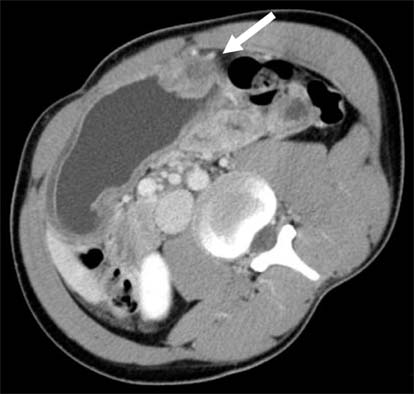
Fig. 2 Abdominal CT shows localized and inhomogeneous enhanced submucosal mass of approximately 3 cm in size on greater curvature of gastric midbody (arrow).

Fig. 3 Endoscopic ultrasonography shows 3-cm- × 2.5-cm-sized heterogeneous submucosal mass on greater curvature of gastric midbody (arrow).
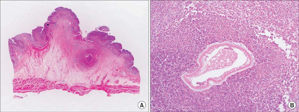
Fig. 4 Histopathological examination shows severe inflammatory cell infiltration, edema, and abscess formation with submucosal eosinophilic granuloma surrounding larva, which are typical findings of gastric anisakiasis (A: H&E, ×1; B: H&E, ×200).
Reference
-
1. van Thiel P, Kuipers FC, Roskam RT. A nematode parasitic to herring, causing acute abdominal syndromes in man. Trop Geogr Med. 1960; 12:97–113.2. Alonso-Gomez A, Moreno-Ancillo A, Lopez-Serrano MC, Suarez-de-Parga JM, Daschner A, Caballero MT, et al. Anisakis simplex only provokes allergic symptoms when the worm parasitises the gastrointestinal tract. Parasitol Res. 2004; 93:378–384.3. Madi LR, Ali M, Legace-Wiens P, Duerksen DR. Gastrointestinal manifestations and management of anisakiasis. Can J Gastroenterol. 2013; 27:126–127.4. Hiramatsu K, Kamiyamamoto S, Ogino H, Satomura Y, Konishi K, Miwa A, et al. A case of acute gastric anisakiasis presenting with malignant tumor-like features: a large gastric vanishing tumor accompanied by local lymph node swelling. Dig Dis Sci. 2004; 49:965–969.5. Takeuchi K, Hanai H, Iida T, Suzuki S, Isobe S. A bleeding gastric ulcer on a vanishing tumor caused by anisakiasis. Gastrointest Endosc. 2000; 52:549–551.6. Ito Y, Ikematsu Y, Yuzawa H, Nishiwaki Y, Kida H, Waki S, et al. Chronic gastric anisakiasis presenting as pneumoperitoneum. Asian J Surg. 2007; 30:67–71.7. Lorenzo S, Iglesias R, Leiro J, Ubeira FM, Ansotegui I, García M, et al. Usefulness of currently available methods for the diagnosis of Anisakis simplex allergy. Allergy. 2000; 55:627–633.8. Kojima K. Parasitic granuloma with special reference to histopathological findings of the Anisakis-like larva infection. Jpn J Parasitol. 1966; 15:30–31.9. Shibata O, Uchida Y, Furusawa T. Acute gastric anisakiasis with special analysis of the location of the worms penetrating the gastric mucosa. In : Ishikura H, Namiki M, editors. Gastric anisakiasis in Japan. Tokyo: Springer-Verlag;1989. p. 53–57.10. Yamazaki M, Hara K, Hasegawa T, Kanazawa M. Vanishing tumor of the stomach? Rinsho Hoshasen. 1976; 21:47–54.
- Full Text Links
-
- Actions
-
Cited
- CITED
-
- Close
- Share
-
- Similar articles
-
- A Case of Gastric Anisakiasis Causing Severe Gastric Ulcer Bleeding
- A Case of Gastric Anisakiasis Presenting with Submucosal Tumor
- A case of acute gastric anisakiasis provoking severe clinical problems by multiple infection
- Three Cases of Coexistence of Gastric Cancer and Duodenal Ulcer
- Study on the Gastric Cancer Initially Diagnosed as Benign Gastric Ulcer during Endoscopic Follow-up
