The Olfactory Groove Schwannoma Attached to the Cribriform Plate: A Case Report
- Affiliations
-
- 1Department of Neurosurgery, National Health Insurance Service Ilsan Hospital, Goyang, Korea. khyang@nhimc.or.kr
- 2Department of Radiology, National Health Insurance Service Ilsan Hospital, Goyang, Korea.
- 3Department of Pathology, National Health Insurance Service Ilsan Hospital, Goyang, Korea.
- KMID: 1882013
- DOI: http://doi.org/10.14791/btrt.2015.3.1.56
Abstract
- The olfactory groove schwannoma is a quite rare tumor. We report a case of a 49-year-old woman with an olfactory groove schwannoma attached to the cribriform plate without olfactory dysfunction. She had no specific neurological symptoms other than a headache, and resection of the tumor showed it to be a schwannoma. About 19 months after the operation, a follow-up MRI showed no evidence of tumor recurrence. Surgical resection through subfrontal approach could be one of the curative modality in managing an olfactory groove schwannoma. An olfactory groove schwannoma should be considered in the differential diagnosis of anterior skull base tumors.
Keyword
MeSH Terms
Figure
-
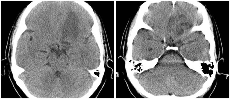
Fig. 1 Computed tomography scans showing a low density lesion in the left frontal lobe.
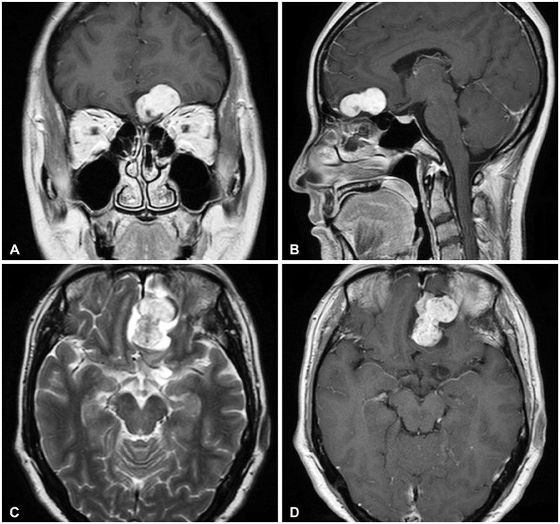
Fig. 2 T1-weighted coronal (A) and sagittal image (B) with gadolinium, showing a solid enhancement of the tumor in the left frontal base. T2-weighted axial MRI revealing an interspersed high signal intensity in the mass, implying cystic component (C). T1-weighted MRI with gadolinium showing a 3 cm sized variegating enhanced mass attached to the cribriform plate (D).
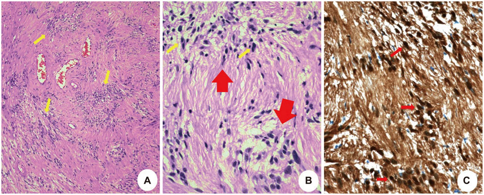
Fig. 3 Pathologic findings. A: On histological examination, the tumor is composed of spindle-shaped cells (arrows) with elongated nuclei and fibrillary cytoplasm (Antoni A pattern), and less cellular areas (Antoni B). B: On high power, Verocay bodies (thick arrows), which are acellular areas surrounded by nuclear palisades composed of spindle cells (thin arrows), the characteristics of schwannoma are well observed (H&E, ×200). C: These spindle cells (arrows) show diffuse and strong immunoreactivity for S-100 protein (×200).
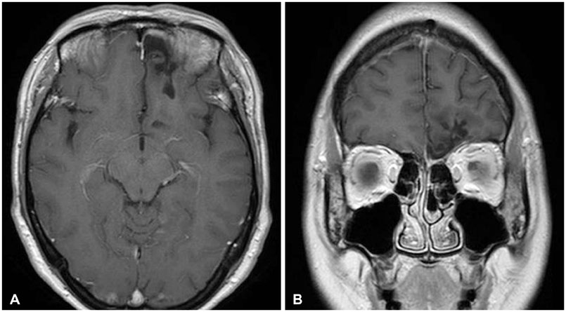
Fig. 4 Post op MRI with gadolinium enhancement. No evidence of local tumor recurrence. An axial view of the T1-weighted gadolinium enhancement (A), a coronal view of T1-weighted gadolinium enhancement (B).

Fig. 5 Follow-up MRI 19 months after operation. No evidence of local tumor recurrence. An axial view of T1-weighted gadolinium enhancement (A), a coronal view of T1-weighted gadolinium enhancement (B).
Cited by 1 articles
-
Subfrontal Schwannoma Extended Broadly to Nasal Cavity Treated by Gamma Knife Radiosurgery Following Surgical Excision: A Case Report
Soo Hee Kim, Jung Hwan Lee, Soon Ki Sung, Chang Hwa Choi
Brain Tumor Res Treat. 2017;5(2):116-119. doi: 10.14791/btrt.2017.5.2.116.
Reference
-
1. Carron JD, Singh RV, Karakla DW, Silverberg M. Solitary schwannoma of the olfactory groove: case report and review of the literature. Skull Base. 2002; 12:163–166.
Article2. Figueiredo EG, Gomes MQ, Soga Y, Amorim RL, Rosemberg S, Teixeira MJ. A rare case of olfactory groove schwannoma. Arq Neuropsiquiatr. 2009; 67:534–535.
Article3. Choi YS, Sung KS, Song YJ, Kim HD. Olfactory schwannoma-case report-. J Korean Neurosurg Soc. 2009; 45:103–106.
Article4. Yasuda M, Higuchi O, Takano S, Matsumura A. Olfactory ensheathing cell tumor: a case report. J Neurooncol. 2006; 76:111–113.
Article5. Shenoy SN, Raja A. Cystic olfactory groove schwannoma. Neurol India. 2004; 52:261–262.6. Li YP, Jiang S, Zhou PZ, Ni YB. Solitary olfactory schwannoma without olfactory dysfunction: a new case report and literature review. Neurol Sci. 2012; 33:137–142.
Article
