Obstet Gynecol Sci.
2013 Jan;56(1):41-44. 10.5468/OGS.2013.56.1.41.
Hepatoid carcinoma of the ovary without staining for alpha-fetoprotein
- Affiliations
-
- 1Department of Obstetrics and Gynecology, Samsung Medical Center, Sungkyunkwan University School of Medicine, Seoul, Korea. bgkim@skku.edu
- KMID: 1857185
- DOI: http://doi.org/10.5468/OGS.2013.56.1.41
Abstract
- Primary hepatoid carcinoma of the ovary (HCO) is a rare type of ovarian tumor that resembles hepatocellular carcinoma both histologically and immunohistochemically in its staining for alpha-fetoprotein (AFP). We describe a 51-year-old woman who presented to our hospital complaining of abdominal pain. Computed tomography scan revealed a large tumor in the pelvis. She underwent total hysterectomy and bilateral salpingo-oophorectomy with tumorectomy. A right ovarian mass measuring 9x8x6 cm was found. Histological diagnosis was hepatoid carcinoma of the right ovary. But, immunohistochemically, tumor cells were not immunoreactive for AFP and there was no elevation of serum AFP level. This is the first report of an ovarian carcinoma with typical histologic features of HCO with negative staining for AFP and normal level of serum AFP in the world.
Keyword
MeSH Terms
Figure
-
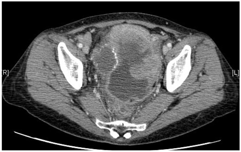
Fig. 1 A computed tomography scan of the abdomen showed 12.5×7.8×11.2 cm sized large heterogeneous pelvic mass invading sigmoid colon and uterus.

Fig. 2 The tumor was composed of solid sheets or aggregates of uniform cells with moredate or abundant eosinophilic cytoplasm, distinct cell borders, and centrally located nuclei with prominent nucleoli (H&E, ×200).

Fig. 3 Immunohistochemistry for alpha-fetoprotein (AFP). The tumor cells are not immunoreactive for AFP (immunostain for AFP, ×200).

Fig. 4 Immunohistochemistry for hepatocyte paraffin-1. The tumor cells are not immunoreactive for hepatocyte paraffin-1 and gonadal dysgenesis was not seen (immunostain for hepatocyte paraffin-1, ×200).
Reference
-
1. Ishikura H, Scully RE. Hepatoid carcinoma of the ovary. A newly described tumor. Cancer. 1987. 60:2775–2784.2. Nishida T, Sugiyama T, Kataoka A, Ushijima K, Ota S, Iwanaga S, et al. Ovarian hepatoid carcinoma without staining for alpha-fetoprotein in the primary site. Int J Gynecol Cancer. 1995. 5:314–318.3. Tochigi N, Kishimoto T, Supriatna Y, Nagai Y, Nikaido T, Ishikura H. Hepatoid carcinoma of the ovary: a report of three cases admixed with a common surface epithelial carcinoma. Int J Gynecol Pathol. 2003. 22:266–271.4. Pandey M, Truica C. Hepatoid carcinoma of the ovary. J Clin Oncol. 2011. 29:e446–e448.5. Kwon JE, Kim SH, Cho NH. No ancillary finding is valid to distinguish a primary ovarian hepatoid carcinoma from metastatic hepatocellular carcinoma. Int J Gynecol Cancer. 2006. 16:1691–1694.6. Choi SM, Park CS, Oh SH, Kim TJ, Song SY, Ahn GH, et al. A case of hepatoid carcinoma of the ovary. Korean J Obstet Gynecol. 2000. 43:141–144.7. Jang KT, Sunwoo JK, Seo SH, Kim CJ, Bae DH. A case of hepatoid carcinoma of the ovary. Korean J Obstet Gynecol. 1996. 39:427–431.8. Trivedi P, Dave K, Shah M, Karelia N, Patel D, Wadhwa M. Hepatoid carcinoma of the ovary: a case report. Eur J Gynaecol Oncol. 1998. 19:167–169.
- Full Text Links
-
- Actions
-
Cited
- CITED
-
- Close
- Share
-
- Similar articles
-
- Primary ovarian hepatoid carcinoma: Case report with review of the literature
- A Case of Hepatoid Carcinoma of the Ovary
- Alpha-Fetoprotein-Producing Carcinoma of the Gallbladder: A case report
- Hepatoid Adenocarcinoma of the Stomach: A Pathologic Analysis of 14 cases
- Two Cases of Recovery of Ovarian Function and Spontaneous Pregnancy in Women Who Were Diagnosed as Premature Ovarian Failure
