Melanosis ilei induced by prolonged charcoal ingestion
- Affiliations
-
- 1Department of Internal Medicine, The Catholic University of Korea School of Medicine, Seoul, Korea. jikim@catholic.ac.kr
- 2Department of Hospital Pathology, The Catholic University of Korea School of Medicine, Seoul, Korea.
- KMID: 1853534
- DOI: http://doi.org/10.4174/jkss.2011.81.1.66
Abstract
- Gastrointestinal melanosis is observed most frequently in the colon it also can develop in the ileum, duodenum and esophagus very rarely. Melanosis ilei was thought that causative materials such as aluminum, magnesium, silicate, titanium and other compounds entered the body through the ingestion of agents. We experienced a case of melanosis in the terminal ileum that a 65-year-old female patient ingested 10 g edible charcoal everyday for 3 years to address symptoms of chronic abdominal pain. In Korea, edible charcoal has been considered to be an effective folk remedy for patients with diarrhea or chronic abdominal pain. In our case, a follow up colonoscopy was performed 3.5 years after the termination of the ingestion of edible charcoal, at which point pigmentation was faded color intensity. In conclusion, it is thought that melanosis ilei is a rare disease by ingestion of causative materials and is discontinuous, local and reversible disease.
Keyword
MeSH Terms
Figure
-
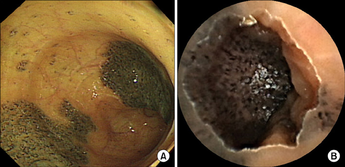
Fig. 1 Colonoscopic features of terminal ileum and capsule endoscopy finding of ileum. (A) The mucosa of the terminal ileum shows punctuate and confluent areas of pigmentation. The lesion is observed as multiple and variable sized geographic black pigmentation spots in the mucosa, and the borders of the lesion are well demarcated. (B) This image shows the well demarcated black pigmented mucosa at terminal ileum.

Fig. 2 Histologic features and electron microscopic finding of melanosis ilei. (A) Light microscope shows macrophages containing black coarse pigments (arrow) in the lamina propria and submucosa (H&E, ×200). (B) This image shows pigment granules at the microvilli and cytoplasms of epithelial cells (arrow) (×4,000).
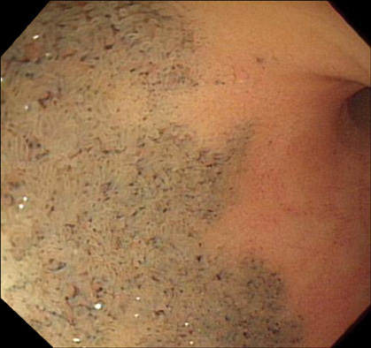
Fig. 3 Follow-up colonoscopy 3.5 years later. The pigment deposition was still observable but had diminished in color intensity.
Reference
-
1. Ghadially FN, Parry EW. An electron-microscope and histochemical study of melanosis coli. J Pathol Bacteriol. 1966. 92:313–317.2. Urbanski SJ, Arsenault AL, Green FH, Haber G. Pigment resembling atmospheric dust in Peyer's patches. Mod Pathol. 1989. 2:222–226.3. Fernando SS. Pseudomelanosis duodeni: a case report with electron-probe X-ray analysis. Pathology. 1990. 22:169–172.4. Yoon JH, Eum SH, Kim SY, Kim CY, Hwang HS, Lee HH, et al. A case of melanosis ilei. Korean J Gastrointest Endosc. 2007. 34:223–226.5. Kim MS, Park YB, Ha BW, Cheung DY, Kim JI, Cho SH, et al. Two cases of melanosis ilei developed after long-standing charcoal ingestion. Korean J Gastrointest Endosc. 2008. 36:36–39.6. Lee TH, Lee SH, Park JH, Park DH, Park JY, Kim HS, et al. Melanosis ilei associated with chronic ingestion of edible charcoal. Gastrointest Endosc. 2008. 67:1174.7. Cha JM, Lee JI, Joo KR, Jung SW, Shin HP. Melanosis ilei associated with chronic ingestion of oral iron. Gut Liver. 2009. 3:315–317.8. Schrodt GR. Melanosis coli: a study with the electron microscope. Dis Colon Rectum. 1963. 6:277–283.9. Ghadially FN, Walley VM. Pigments of the gastrointestinal tract: a comparison of light microscopic and electron microscopic findings. Ultrastruct Pathol. 1995. 19:213–219.10. Susaya J, Kim KH, Ahn JW, Jung MC, Kang CH. BBQ charcoal combustion as an important source of trace metal exposure to humans. J Hazard Mater. 2010. 176:932–937.
