Silent Microembolism on Diffusion-Weighted MRI after Coil Embolization of Cerebral Aneurysms
- Affiliations
-
- 1Department of Neurosurgery, Seoul Paik Hospital, Inje University College of Medicine, Seoul, Korea.
- 2Department of Neurosurgery, Seoul St. Mary's Hospital, The Catholic University of Korea, Seoul, Korea. nsshin@catholic.ac.kr
- KMID: 1841093
- DOI: http://doi.org/10.5469/neuroint.2012.7.2.77
Abstract
- PURPOSE
The purpose of this study was to investigate the frequency and risk factors of procedure-related thromboembolism on diffusion-weighted imaging (DWI) associated with aneurysmal coil embolization.
MATERIALS AND METHODS
We prospectively evaluated 39 consecutive patients with a cerebral aneurysm with DWI after coil embolization. All hyperintense lesions on DWI with a drop of apparent diffusion coefficient values were classified into acute thromboembolic infarction (larger than 5 mm in maximal diameters, and located in the vascular territory of the parent artery) and silent microembolism (single or multiple tiny dot-like lesion, less than 5 mm, usually 1-2 mm in size). Possible risk factors for thromboembolic events included vascular risk factors, aneurysmal factors, and procedure-related factors.
RESULTS
Hyperintense lesions on DWI were seen in 17 (43.6%) patients and symptomatic DWI positive lesions were four (10.3%). Acute thromboembolic infarction was observed in seven (17.9%) patients and silent microembolism in 14 (35.9%) patients. Numbers of silent microembolism ranged from 1 to 15 (mean: 2.86, standard deviation: 3.74). Silent microembolisms were located at ipsilateral (n=3, 21.4%), contralateral (n=5, 35.7%), bilateral (n=4, 28.6%), and not related (n=2, 14.3%) to the procedure site. There were no statistical significant risk factors in acute thromboembolic infarction. However, incidence of silent microembolisms was significantly correlated with left side approach (odds ratio, 4.44, 95% confidence interval, 1.08-18.36; P=0.03).
CONCLUSION
Left side approach may have increased the likelihood of asymptomatic multiple scattered microemboli after aneurysmal coiling procedures. Particular care must be taken in the handling of guiding catheters, especially when proving left side great vessels.
Keyword
MeSH Terms
Figure
-
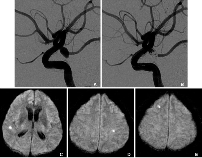
Fig. 1 A 55-year-old male with a ruptured posterior communicating artery (PComA) aneurysm.A. Preoperative left internal carotid artery lateral angiograms.B. Immediate postembolization image shows partial aneurysmal occlusion with a neck remnant around the right PComA orifices.C-E. Diffusion-weighted images obtained 24 hours after the procedure shows multiple small hyperintense signals on ipsilateral and contralateral side of the procedures without any symptom.
Cited by 2 articles
-
Thromboembolic Event Detected by Diffusion Weighted Magnetic Resonance Imaging After Coil Embolization of Cerebral Aneurysms
Dong-Ho Seo, Seok-Mann Yoon, Hye-Ran Park, Jai-Joon Shim, Hack-Gun Bae, Il-Gyu Yun
J Cerebrovasc Endovasc Neurosurg. 2014;16(3):175-183. doi: 10.7461/jcen.2014.16.3.175.Microembolism after Endovascular Treatment of Unruptured Cerebral Aneurysms: Reduction of its Incidence by Microcatheter Lumen Aspiration
Dae Yoon Kim, Jung Cheol Park, Jae Kyun Kim, Yu Sub Sung, Eun Suk Park, Jae Hyuk Kwak, Choong-Gon Choi, Deok Hee Lee
Neurointervention. 2015;10(2):67-73. doi: 10.5469/neuroint.2015.10.2.67.
Reference
-
1. Guglielmi G, Vinuela F, Sepetka I, Macellari V. Electrothrombosis of saccular aneurysms via endovascular approach. Part 1: Electrochemical basis, technique, and experimental results. J Neurosurg. 1991; 75:1–7. PMID: 2045891.2. Guglielmi G, Vinuela F, Dion J, Duckwiler G. Electrothrombosis of saccular aneurysms via endovascular approach. Part 2: Preliminary clinical experience. J Neurosurg. 1991; 75:8–14. PMID: 2045924.3. Biondi A, Oppenheim C, Vivas E, Casasco A, Lalam T, Sourour N, et al. Cerebral aneurysms treated by Guglielmi detachable coils: evaluation with diffusion-weighted MR imaging. AJNR Am J Neuroradiol. 2000; 21:957–963. PMID: 10815677.4. Derdeyn CP, Cross DT 3rd, Moran CJ, Brown GW, Pilgram TK, Diringer MN, et al. Postprocedure ischemic events after treatment of intracranial aneurysms with Guglielmi detachable coils. J Neurosurg. 2002; 96:837–843. PMID: 12008697.
Article5. Pelz DM, Lownie SP, Fox AJ. Thromboembolic events associated with the treatment of cerebral aneurysms with Guglielmi detachable coils. AJNR Am J Neuroradiol. 1998; 19:1541–1547. PMID: 9763391.6. Rordorf G, Bellon RJ, Budzik RE Jr, Farkas J, Reinking GF, Pergolizzi RS, et al. Silent thromboembolic events associated with the treatment of unruptured cerebral aneurysms by use of Guglielmi detachable coils: prospective study applying diffusion-weighted imaging. AJNR Am J Neuroradiol. 2001; 22:5–10. PMID: 11158880.7. Soeda A, Sakai N, Murao K, Sakai H, Ihara K, Yamada N, et al. Thromboembolic events associated with Guglielmi detachable coil embolization with use of diffusion-weighted MR imaging. Part II. Detection of the microemboli proximal to cerebral aneurysm. AJNR Am J Neuroradiol. 2003; 24:2035–2038. PMID: 14625228.8. Soeda A, Sakai N, Sakai H, Iihara K, Yamada N, Imakita S, et al. Thromboembolic events associated with Guglielmi detachable coil embolization of asymptomatic cerebral aneurysms. Evaluation of 66 consecutive cases with use of diffusion-weighted MR imaging. AJNR Am J Neuroradiol. 2003; 24:127–132. PMID: 12533341.9. Albayram S, Selcuk H, Kara B, Bozdag E, Uzma O, Kocer N, et al. Thromboembolic events associated with balloon-assisted coil embolization: evaluation with diffusion-weighted MR imaging. AJNR Am J Neuroradiol. 2004; 25:1768–1777. PMID: 15569744.10. Brooks NP, Turk AS, Niemann DB, Aagaard-Kienitz B, Pulfer K, Cook T. Frequency of thromboembolic events associated with endovascular aneurysm treatment: retrospective case series. J Neurosurg. 2008; 108:1095–1100. PMID: 18518710.
Article11. Fisher CM, Kistler JP, Davis JM. Relation of cerebral vasospasm to subarachnoid hemorrhage visualized by computerized tomographic scanning. Neurosurgery. 1980; 6:1–9. PMID: 7354892.
Article12. Vinuela F, Duckwiler G, Mawad M. Guglielmi detachable coil embolization of acute intracranial aneurysm: perioperative anatomical and clinical outcome in 403 patients. J Neurosurg. 1997; 86:475–482. PMID: 9046305.13. Martin D, Rodesch G, Alvarez H, Lasjaunias P. Preliminary results of embolisation of nonsurgical intracranial aneurysms with GD coils: the 1st year of their use. Neuroradiology. 1996; 38(Suppl 1):S142–S150. PMID: 8811702.
Article14. Bendszus M, Koltzenburg M, Burger R, Warmuth-Metz M, Hofmann E, Solymosi L. Silent embolism in diagnostic cerebral angiography and neurointerventional procedures: a prospective study. Lancet. 1999; 354:1594–1597. PMID: 10560674.
Article15. Moret J, Cognard C, Weill A, Castaings L, Rey A. The "Remodeling technique" in the treatment of wide neck intracranial aneurysms. Angiographic results and clinical follow-up in 56 cases. Interv Neuroradiol. 1997; 3:21–35. PMID: 20678369.16. Malek AM, Halbach VV, Phatouros CC, Lempert TE, Meyers PM, Dowd CF, et al. Balloon-assist technique for endovascular coil embolization of geometrically difficult intracranial aneurysms. Neurosurgery. 2000; 46:1397–1406. PMID: 10834645.
Article17. Nelson PK, Levy DI. Balloon-assisted coil embolization of wide-necked aneurysms of the internal carotid artery: medium-term angiographic and clinical follow-up in 22 patients. AJNR Am J Neuroradiol. 2001; 22:19–26. PMID: 11158882.18. Cloft HJ, Joseph GJ, Dion JE. Risk of cerebral angiography in patients with subarachnoid hemorrhage, cerebral aneurysm, and arteriovenous malformation: a meta-analysis. Stroke. 1999; 30:317–320. PMID: 9933266.
- Full Text Links
-
- Actions
-
Cited
- CITED
-
- Close
- Share
-
- Similar articles
-
- Thromboembolic Events after Coil Embolization of Cerebral Aneurysms: Prospective Study with Diffusion-Weighted Magnetic Resonance Imaging Follow-up
- Recent Trends in the Treatment of Cerebral Aneurysms: Comparison between Endovascular Coil Embolization and Surgical Clipping
- Microembolism after Endovascular Treatment of Unruptured Cerebral Aneurysms: Reduction of its Incidence by Microcatheter Lumen Aspiration
- Endovascular Treatment of Cerebral Aneurysms: Technical Options in Coil Embolization
- Single-session Coil Embolization of Multiple Intracranial Aneurysms
