Comparison of Enterprise and Neuroform Stent-Assisted Coil Embolization of Distal Internal Carotid Artery Aneurysms: Midterm Results from a Single-Center Experience
- Affiliations
-
- 1Department of Radiology, Research Institute for Convergence of Biomedical Science and Technology, Pusan National University Yangsan Hospital, Pusan National University School of Medicine, Yangsan, Korea. skbaik9@gmail.com
- 2Department of Neurosurgery, Research Institute for Convergence of Biomedical Science and Technology, Pusan National University Yangsan Hospital, Pusan National University School of Medicine, Yangsan, Korea.
- KMID: 1839429
- DOI: http://doi.org/10.3348/jksr.2014.70.3.181
Abstract
- PURPOSE
To compare the mid-term follow-up angiographic findings in distal internal carotid artery (ICA) aneurysms treated by stent-assisted coil embolization using the Enterprise or Neuroform stent.
MATERIALS AND METHODS
We included 68 patients with 70 aneurysms: 31 cases with Enterprise and 39 cases with Neuroform. Inclusion criteria were 1) location of the stent within the distal ICA, including the carotid siphon; 2) follow-up angiogram after > 6 months, and 3) single use of the stent for 1 parent artery.
RESULTS
The patients' mean age was 54.9 years (16 male and 52 female). Mean follow-up duration was 9.1 months. At follow-up, there were intraluminal filling defects of the parent artery in 19.4% of the Enterprise group and no filling defect in the Neuroform group. There was no significant in-stent stenosis in either group. Straightening of the parent artery was seen in 35.5% of the Enterprise group and 20.5% of the Neuroform group. Two Enterprise cases showed delayed migration.
CONCLUSION
The Enterprise showed statistically significant intraluminal filling defects of the parent artery compared with the Neuroform. The rates of significant in-stent stenosis and straightening of the parent artery were not significantly different between the Enterprise and the Neuroform groups.
MeSH Terms
Figure
-
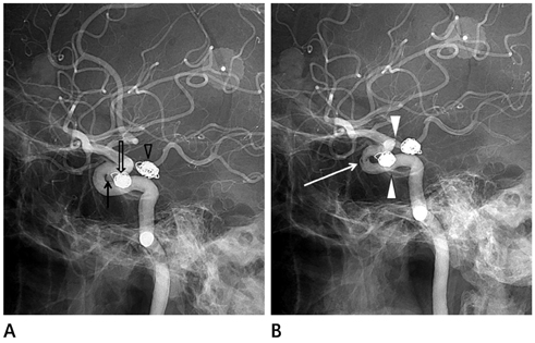
Fig. 1 Intraluminal filling defect (incomplete stent apposition). A. A 67-year-old woman has a left P-com aneurysm (arrowhead) and two right paraclinoid aneurysms (arrow and open arrow). Immediate post-procedural angiography shows right paraclinoid ICA aneurysm after stent-assisted coiling using a 4.5 × 28 mm Enterprise stent with complete occlusion and a just proximal paraclinoid aneurysm treated by stent only. B. Eight-month follow-up angiography reveals an intraluminal filling defect (arrow) at the substented area of the siphon (between proximal arrowhead and distal arrowhead). Note.-ICA = internal carotid artery
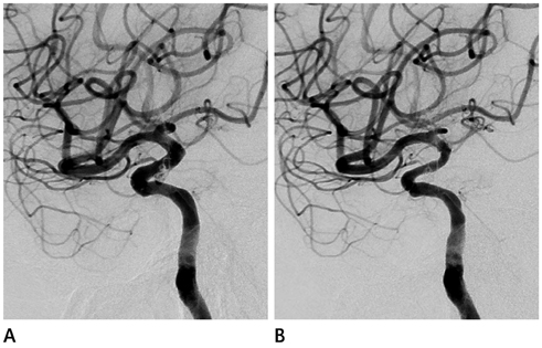
Fig. 2 In-stent stenosis. A 64-year-old woman with an incidentally found aneurysm. A. Immediate post-procedural angiography shows left paraclinoid ICA aneurysm after stent-assisted coiling using a 4.5 × 20 mm Neuroform stent. Subtle contrast filling of the aneurysm sac is seen. B. Six-month follow-up angiography shows that the aneurysm underwent progressive occlusion as there is no residual flow visible. In addition, there is mild (40%) narrowing of substented area of the parent artery. But it is not significant in-stent stenosis, according to our definition: we defined the term "significant in-stent stenosis" as more than 50% narrowing within the stent-deployment area of the parent artery. Note.-ICA = internal carotid artery
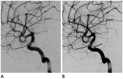
Fig. 3 Parent artery straightening. A 36-year-old woman presented with severe headache. Examination revealed subarachnoid hemorrhage due to ruptured aneurysm. A. Immediate post-procedural angiography shows a right paraclinoid ICA aneurysm after stent-assisted coiling using a 4.5 × 28 mm Enterprise stent with complete occlusion. B. Twelve-month follow-up angiography reveals increased vascular curvature of the carotid siphon ("straightening") and focal intraluminal filling defect at the substented area (arrow). Note.-ICA = internal carotid artery
Reference
-
1. Huang QH, Wu YF, Xu Y, Hong B, Zhang L, Liu JM. Vascular geometry change because of endovascular stent placement for anterior communicating artery aneurysms. AJNR Am J Neuroradiol. 2011; 32:1721–1725.2. Molyneux AJ, Kerr RS, Birks J, Ramzi N, Yarnold J, Sneade M, et al. Risk of recurrent subarachnoid haemorrhage, death, or dependence and standardised mortality ratios after clipping or coiling of an intracranial aneurysm in the International Subarachnoid Aneurysm Trial (ISAT): long-term follow-up. Lancet Neurol. 2009; 8:427–433.3. Wakhloo AK, Linfante I, Silva CF, Samaniego EA, Dabus G, Etezadi V, et al. Closed-cell stent for coil embolization of intracranial aneurysms: clinical and angiographic results. AJNR Am J Neuroradiol. 2012; 33:1651–1656.4. Lavine SD, Larsen DW, Giannotta SL, Teitelbaum GP. Parent vessel Guglielmi detachable coil herniation during wide-necked aneurysm embolization: treatment with intracranial stent placement: two technical case reports. Neurosurgery. 2000; 46:1013–1017.5. Phatouros CC, Sasaki TY, Higashida RT, Malek AM, Meyers PM, Dowd CF, et al. Stent-supported coil embolization: the treatment of fusiform and wide-neck aneurysms and pseudoaneurysms. Neurosurgery. 2000; 47:107–113. discussion 113-115.6. Gallas S, Pasco A, Cottier JP, Gabrillargues J, Drouineau J, Cognard C, et al. A multicenter study of 705 ruptured in tracranial aneurysms treated with Guglielmi detachable coils. AJNR Am J Neuroradiol. 2005; 26:1723–1731.7. Heller RS, Malek AM. Parent vessel size and curvature strongly influence risk of incomplete stent apposition in enterprise intracranial aneurysm stent coiling. AJNR Am J Neuroradiol. 2011; 32:1714–1720.8. Krischek O, Miloslavski E, Fischer S, Shrivastava S, Henkes H. A comparison of functional and physical properties of self-expanding intracranial stents [Neuroform3, Wingspan, Solitaire, Leo+, Enterprise]. Minim Invasive Neurosurg. 2011; 54:21–28.9. Gao B, Malek AM. Possible mechanisms for delayed migration of the closed cell--designed enterprise stent when used in the adjunctive treatment of a basilar artery aneurysm. AJNR Am J Neuroradiol. 2010; 31:E85–E86.10. Ebrahimi N, Claus B, Lee CY, Biondi A, Benndorf G. Stent conformity in curved vascular models with simulated aneurysm necks using flat-panel CT: an in vitro study. AJNR Am J Neuroradiol. 2007; 28:823–829.11. Lubicz B, François O, Levivier M, Brotchi J, Balériaux D. Preliminary experience with the enterprise stent for endovascular treatment of complex intracranial aneurysms: potential advantages and limiting characteristics. Neurosurgery. 2008; 62:1063–1069. discussion 1069-1070.12. Yang PF, Liu JM, Huang QH, Zhao WY, Hong B, Xu Y, et al. Preliminary experience and short-term follow-up results of treatment of wide-necked or fusiform cerebral aneurysms with a self-expanding, closed-cell, retractable stent. J Clin Neurosci. 2010; 17:837–841.13. Raymond J, Roy D, Bojanowski M, Moumdjian R, L'Espérance G. Endovascular treatment of acutely ruptured and unruptured aneurysms of the basilar bifurcation. J Neurosurg. 1997; 86:211–219.14. van Swieten JC, Koudstaal PJ, Visser MC, Schouten HJ, van Gijn J. Interobserver agreement for the assessment of handicap in stroke patients. Stroke. 1988; 19:604–607.15. Maldonado IL, Machi P, Costalat V, Mura T, Bonafé A. Neuroform stent-assisted coiling of unruptured intracranial aneurysms: short- and midterm results from a single-center experience with 68 patients. AJNR Am J Neuroradiol. 2011; 32:131–136.16. Turk AS, Niemann DB, Ahmed A, Aagaard-Kienitz B. Use of self-expanding stents in distal small cerebral vessels. AJNR Am J Neuroradiol. 2007; 28:533–536.17. Heller RS, Miele WR, Do-Dai DD, Malek AM. Crescent sign on magnetic resonance angiography revealing incomplete stent apposition: correlation with diffusion-weighted changes in stent-mediated coil embolization of aneurysms. J Neurosurg. 2011; 115:624–632.18. Piotin M, Blanc R, Spelle L, Mounayer C, Piantino R, Schmidt PJ, et al. Stent-assisted coiling of intracranial aneurysms: clinical and angiographic results in 216 consecutive aneurysms. Stroke. 2010; 41:110–115.19. Rathore S, Terashima M, Habara M, Kinoshita Y, Nasu K, Katoh O, et al. Incomplete stent apposition after coronary stent implantation: myth or reality? J Interv Cardiol. 2009; 22:341–349.20. Bennett MR. Vascular pathology as a result of drug-eluting stents. Heart. 2007; 93:895–896.21. du Mesnil de Rochemont R, Yan B, Zanella FE, Rüfenacht DA, Berkefeld J. Conformability of balloon-expandable stents to the carotid siphon: an in vitro study. AJNR Am J Neuroradiol. 2006; 27:324–326.22. Ozaki Y, Okumura M, Ismail TF, Naruse H, Hattori K, Kan S, et al. The fate of incomplete stent apposition with drug-eluting stents: an optical coherence tomography-based natural history study. Eur Heart J. 2010; 31:1470–1476.23. King RM, Chueh JY, vanderBom IM, Silva CF, Carniato SL, Spilberg G, et al. The effect of intracranial stent implantation on the curvature of the cerebrovasculature. AJNR Am J Neuroradiol. 2012; 33:1657–1662.24. Gao B, Baharoglu MI, Cohen AD, Malek AM. Stent-assisted coiling of intracranial bifurcation aneurysms leads to immediate and delayed intracranial vascular angle remodeling. AJNR Am J Neuroradiol. 2012; 33:649–654.25. Yoon KW, Kim YJ. In-stent stenosis of stent assisted endovascular treatment on intracranial complex aneurysms. J Korean Neurosurg Soc. 2010; 48:485–489.26. Kipshidze N, Dangas G, Tsapenko M, Moses J, Leon MB, Kutryk M, et al. Role of the endothelium in modulating neointimal formation: vasculoprotective approaches to attenuate restenosis after percutaneous coronary interventions. J Am Coll Cardiol. 2004; 44:733–739.27. Wakhloo AK, Mandell J, Gounis MJ, Brooks C, Linfante I, Winer J, et al. Stent-assisted reconstructive endovascular repair of cranial fusiform atherosclerotic and dissecting aneurysms: long-term clinical and angiographic follow-up. Stroke. 2008; 39:3288–3296.28. Marks MP, Wojak JC, Al-Ali F, Jayaraman M, Marcellus ML, Connors JJ, et al. Angioplasty for symptomatic intracranial stenosis: clinical outcome. Stroke. 2006; 37:1016–1020.29. Fiorella D, Albuquerque FC, Woo H, Rasmussen PA, Masaryk TJ, McDougall CG. Neuroform in-stent stenosis: incidence, natural history, and treatment strategies. Neurosurgery. 2006; 59:34–42. discussion 34-42.30. Kearney M, Pieczek A, Haley L, Losordo DW, Andres V, Schainfeld R, et al. Histopathology of in-stent restenosis in patients with peripheral artery disease. Circulation. 1997; 95:1998–2002.31. Hoffmann R, Mintz GS, Dussaillant GR, Popma JJ, Pichard AD, Satler LF, et al. Patterns and mechanisms of in-stent restenosis. A serial intravascular ultrasound study. Circulation. 1996; 94:1247–1254.32. McLaughlin N, McArthur DL, Martin NA. Use of stent-assisted coil embolization for the treatment of wide-necked aneurysms: a systematic review. Surg Neurol Int. 2013; 4:43.33. Rodriguez GJ, Maud A, Taylor RA. Another delayed migration of an enterprise stent. AJNR Am J Neuroradiol. 2009; 30:E57.34. Kelly ME, Turner RD 4th, Moskowitz SI, Gonugunta V, Hussain MS, Fiorella D. Delayed migration of a self-expanding intracranial microstent. AJNR Am J Neuroradiol. 2008; 29:1959–1960.35. Lavine SD, Meyers PM, Connolly ES, Solomon RS. Spontaneous delayed proximal migration of enterprise stent after staged treatment of wide-necked basilar aneurysm: technical case report. Neurosurgery. 2009; 64:E1012. discussion E1012.36. Izar B, Rai A, Raghuram K, Rotruck J, Carpenter J. Comparison of devices used for stent-assisted coiling of intracranial aneurysms. PLoS One. 2011; 6:e24875.37. Sedat J, Chau Y, Mondot L, Vargas J, Szapiro J, Lonjon M. Endovascular occlusion of intracranial wide-necked aneurysms with stenting (Neuroform) and coiling: mid-term and long-term results. Neuroradiology. 2009; 51:401–409.38. Biondi A, Janardhan V, Katz JM, Salvaggio K, Riina HA, Gobin YP. Neuroform stent-assisted coil embolization of wide-neck intracranial aneurysms: strategies in stent deployment and midterm follow-up. Neurosurgery. 2007; 61:460–468. discussion 468-469.
- Full Text Links
-
- Actions
-
Cited
- CITED
-
- Close
- Share
-
- Similar articles
-
- Safety and effectiveness of Neuroform Atlas stent-assisted coil embolization for ruptured intracranial aneurysms
- In-Stent Stenosis of Stent-Assisted Coil Embolization of the Supraclinoid Internal Carotid Artery Aneurysm
- Usefulness of Silent MRA for Evaluation of Aneurysm after Stent-Assisted Coil Embolization
- Dual Stent-Assisted Coil Embolization for Fusiform Aneurysm Arising From Persistent Trigeminal Artery
- Initial Experience with Neuroform EZ in the Treatment of Wide-neck Cerebral Aneurysms
