Ultrasound and CT Findings of Primary Leiomyosarcoma in the Gallbladder: A Case Report
- Affiliations
-
- 1Department of Radiology, Wonkwang University School of Medicine and Hospital, Iksan, Korea. yjyh@wonkwang.ac.kr
- 2Department of Radiology, Iksan Hospital, Iksan, Korea.
- 3Department of Surgery, Wonkwang University School of Medicine and Hospital, Iksan, Korea.
- 4Department of Pathology, Wonkwang University School of Medicine and Hospital, Iksan, Korea.
- KMID: 1839422
- DOI: http://doi.org/10.3348/jksr.2014.70.2.133
Abstract
- Leiomyosarcoma of the gallbladder is a very rare subgroup for gallbladder sarcoma. Herein, we report the ultrasound, computed tomography and positron emission tomography-computed tomography imaging findings on a case of primary leiomyosarcoma of the gallbladder. Abdominal ultrasonography indicates a heterogeneous hyperechoic submucosal mass with hypervascularity and displacement of overlying mucosal layers by the mass. Computed tomography reveal that the tumor is a well-defined and heterogeneously enhancing solid mass with overlying thick mucosal layers. Positron emission tomography-computed tomography visualizes the large gallbladder mass as a hypermetabolic lesion.
Figure
-
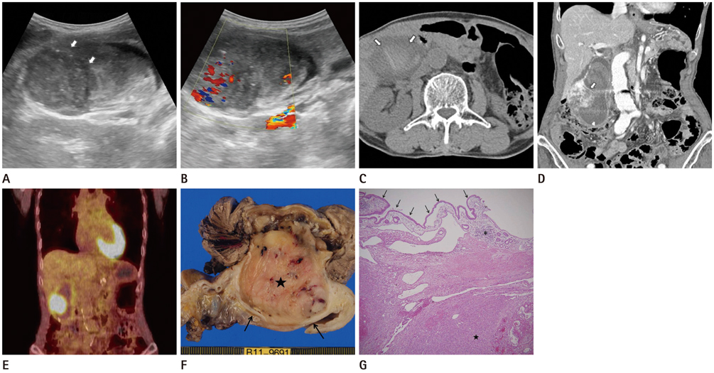
Fig. 1 An 82-year-old male patient with incidentally detected GB mass on ultrasonography. A. Transabdominal ultrasonography demonstrates a large intraluminal protruding solid mass with heterogeneous echogenecity and thickened overlying echogenic mucosa (arrows). Hyperechoic hematoma is also shown beside the mass, within the GB lumen. B. Color Doppler ultrasonography reveals hypervascularity inside the mass. C. Noncontrast axial CT image shows hyperattenuating hematoma within the GB lumen (arrows). D. Coronal reformation image of contrast enhanced CT shows a large polypoid mass displacing the thick enhancing mucosal layer (arrow). It shows heterogeneous enhancement because of multiple necrotic and hemorrhagic foci. E. The GB mass is demonstrated as a marked hypermetabolic lesion on PET-CT image (SUV = 8.3). F. Gross pathologic specimen demonstrates a large well defined solid mass (★) arising from wall of the GB and thick overlying mucosal layer (arrows). This mass containes some necrotic and hemorrhagic foci. G. Photomicrograph (H&E stain, × 40) shows a leiomyosarcoma (★) of the GB, which is located below the mucosa as a single layer of columnar epithelium (black arrows) with an underlying lamina propria, irregular muscle layer, and loose perimuscular connective tissue (*). Note.-GB = gallbladder, PET-CT = positron emission tomography-CT, SUV = standardized uptake value
Reference
-
1. Bernardos L, Trujillo A, Huete A, Colon A, Martínez D, Calleja J, et al. [Primary leiomyosarcoma of the gallbladder]. Rev Esp Enferm Dig. 2004; 96:286–287.2. Willén R, Willén H. Primary sarcoma of the gallbladder. A light and electronmicroscopical study. Virchows Arch A Pathol Anat Histol. 1982; 396:91–102.3. Savlania A, Behera A, Vaiphei K, Singh H, Dhiman RK, Duseja A, et al. Primary leiomyosarcoma of gallbladder: a rare diagnosis. Case Rep Gastrointest Med. 2012; 2012:287012.4. Newmark H 3rd, Kliewer K, Curtis A, DenBesten L, Enenstein W. Primary leiomyosarcoma of gallbladder seen on computed tomography and ultrasound. Am J Gastroenterol. 1986; 81:202–204.5. Danikas D, Theodorou SJ, Singh R, Camal DE. Leiomyosarcoma of the gallbladder: a case report. Am Surg. 2001; 67:873–874.6. Vaittinen E. Sarcoma of the gall-bladder. Ann Chir Gynaecol Fenn. 1972; 61:185–189.7. Kim MJ, Kim KW, Kim HC, Kim SY, Park SH, Kim AY, et al. Unusual malignant tumors of the gallbladder. AJR Am J Roentgenol. 2006; 187:473–480.8. Coelho JC, Wallbach A, Kasting G, Moreira RR. Ultrasonic diagnosis of primary sarcoma of the gallbladder. J Clin Ultrasound. 1984; 12:168–170.9. Guo KJ, Yamaguchi K, Enjoji M. Undifferentiated carcinoma of the gallbladder. A clinicopathologic, histochemical, and immunohistochemical study of 21 patients with a poor prognosis. Cancer. 1988; 61:1872–1879.10. Ishii T, Watanabe K, Kin H, Muro M, Uda K, Idani H, et al. A case of primary leiomyosarcoma of the gallbladder synchronously overlapped with early gastric cancer. Jpn J Gastroenterol Surg. 2002; 35:517–521.
- Full Text Links
-
- Actions
-
Cited
- CITED
-
- Close
- Share
-
- Similar articles
-
- Double Primary Cancer of the Gallbladder and Cystic Duct: A Case Report
- CT and MR Imaging Findings of Primary Leiomyosarcoma of the Vagina: A Case Report
- Imaging Findings of Primary Adrenal Leiomyosarcoma: A Case Report
- Primary leiomyosarcoma of gallbladder
- Primary Leiomyosarcoma of the Breast: A Case Report
