J Korean Surg Soc.
2012 Dec;83(6):403-407. 10.4174/jkss.2012.83.6.403.
Primary leiomyosarcoma of gallbladder
- Affiliations
-
- 1Department of Surgery, School of Medicine, Pusan National University, Biomedical Research Institute, Pusan National University Hospital, Busan, Korea. seohi71@hanmail.net
- 2Department of Radiology, School of Medicine, Pusan National University, Biomedical Research Institute, Pusan National University Hospital, Busan, Korea.
- 3Department of Pathology, School of Medicine, Pusan National University, Biomedical Research Institute, Pusan National University Hospital, Busan, Korea.
- KMID: 1437544
- DOI: http://doi.org/10.4174/jkss.2012.83.6.403
Abstract
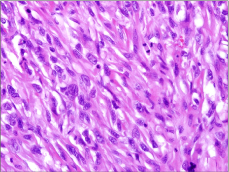
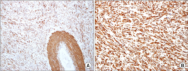
- Malignant mesenchymalneoplasms of the gallbladder are extremely rare with only 105 cases of primary gallbladder sarcoma having been described. It has a very aggressive behavior and is usually diagnosed at advanced stages. Therefore, curative surgical management may not be possible. We performed a radical cholecystectomy (S4b + S5 segmentectomy), omentectomy and small bowel resection in a 54-year-old patient with locally invasive leiomyosarcoma of the gallbladder. Further studies are needed to confirm the benefit of aggressive treatment for patients with leiomyosarcoma of the gallbladder.
Keyword
Figure
Reference
-
1. Albores-Saavera J, Henson DE, Klimstra DS. Atlas of tumor pathology. 3rd series, fascicle 27. Tumors of the gallbladder, and extrahepatic bile duct and ampulla of water. 2000. Washington DC: Armed Forces Institute of Pathology.2. Husain EA, Prescott RJ, Haider SA, Al-Mahmoud RW, Zelger BG, Zelger B, et al. Gallbladder sarcoma: a clinicopathological study of seven cases from the UK and Austria with emphasis on morphological subtypes. Dig Dis Sci. 2009. 54:395–400.3. Newmark H 3rd, Kliewer K, Curtis A, DenBesten L, Enenstein W. Primary leiomyosarcoma of gallbladder seen on computed tomography and ultrasound. Am J Gastroenterol. 1986. 81:202–204.4. Sawan AS, Salama SI. Leiomyosarcoma of the gallbladder. J King Abdulaziz Univ Med Sci. 2010. 17:80–88.5. Anaya DA, Lev DC, Pollock RE. The role of surgical margin status in retroperitoneal sarcoma. J Surg Oncol. 2008. 98:607–610.6. Zalupski M, Metch B, Balcerzak S, Fletcher WS, Chapman R, Bonnet JD, et al. Phase III comparison of doxorubicin and dacarbazine given by bolus versus infusion in patients with soft-tissue sarcomas: a Southwest Oncology Group study. J Natl Cancer Inst. 1991. 83:926–932.7. Grobmyer SR, Maki RG, Demetri GD, Mazumdar M, Riedel E, Brennan MF, et al. Neo-adjuvant chemotherapy for primary high-grade extremity soft tissue sarcoma. Ann Oncol. 2004. 15:1667–1672.8. Adjuvant chemotherapy for localised resectable soft-tissue sarcoma of adults: meta-analysis of individual data. Sarcoma Meta-analysis Collaboration. Lancet. 1997. 350:1647–1654.9. Antman KH, Elias A. Dana-Farber Cancer Institute studies in advanced sarcoma. Semin Oncol. 1990. 17:1 Suppl 2. 7–15.10. Fotiadis C, Gugulakis A, Nakopoulou L, Sechas M. Primary leiomyosarcoma of the gallbladder. Case report and review of the literature. HPB Surg. 1990. 2:211–214.
- Full Text Links
- Actions
-
Cited
- CITED
-
- Close
- Share
- Similar articles
-
- Ultrasound and CT Findings of Primary Leiomyosarcoma in the Gallbladder: A Case Report
- Imaging Findings of Primary Adrenal Leiomyosarcoma: A Case Report
- A Case of Primary Leiomyosarcoma of the Lung
- Two Cases of Primary Uterine Leiomyosarcoma:MR Findings and Pathologic Correlation
- A case of primary pulmonary leiomyosarcoma

