A Case of Intra- and Extra-Mural Hematomas During Recanalization for Chronic Total Occlusion
- Affiliations
-
- 1Division of Cardiology, Department of Internal Medicine, Keimyung University Dongsan Medical Center, Daegu, Korea. shur@dsmc.or.kr
- KMID: 1826180
- DOI: http://doi.org/10.4070/kcj.2010.40.11.596
Abstract
- An intramural hematoma is an accumulation of blood between the internal and external elastic membranes within the medial space, whereas an extramural hematoma is a dilution and/or dissemination of blood throughout the adventitia. Intra- and extra-hematomas are observed by intravascular ultrasound during percutaneous coronary intervention (PCI). The patient described herein presented with angina pectoris. Her coronary angiogram showed diffuse narrowing of the mid-left anterior descending artery and total occlusion of the distal right coronary artery (RCA). Intra- and extra-mural hematomas developed during PCI of the RCA; however, the lesions were covered successfully using long drug-eluting stents.
Keyword
MeSH Terms
Figure
-
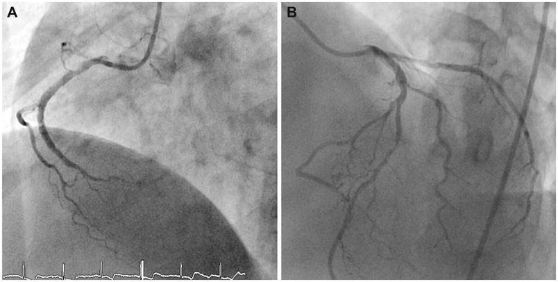
Fig. 1 Diagnostic coronary angiogram revealed total occlusion of the distal RCA (A) and collateral flow from the left coronary artery system to the distal RCA (B). RCA: right coronary artery.
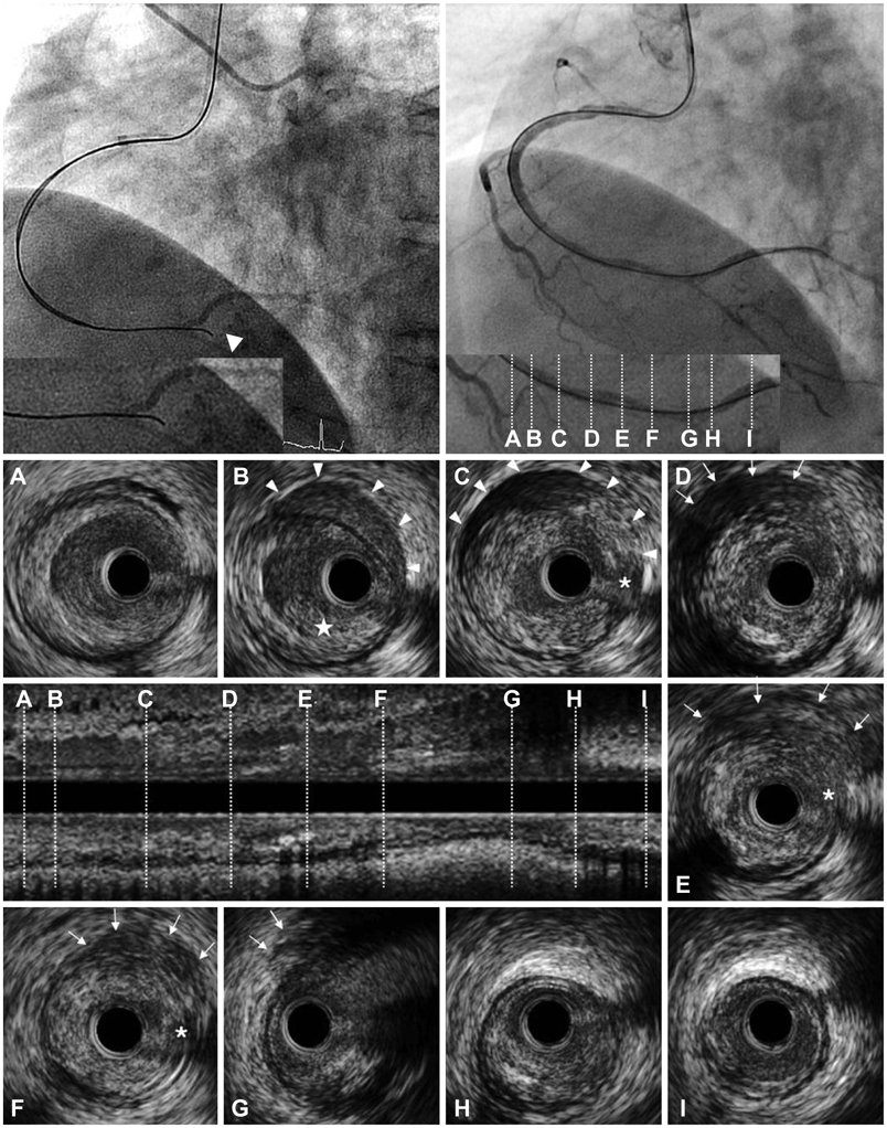
Fig. 2 After advancement of a Conquest Pro guidewire (Asahi, Seto, Japan) using a parallel guidewire technique to the RCA, a coronary angiogram showed that the tip of the guidewire was placed outside of the vessel (arrowhead; upper left). After several attempts to cross the CTO lesions, a Conquest Pro guidewire was succeeded to enter into the true lumen of the PL branch (upper right). The lower panel shows cross-sectional images, as well as a longitudinal image of the IVUS from the PL branch to the distal RCA. A and B: at the proximal reference (in the distal RCA) of the CTO, an intraluminal thrombus (★) and intramural hematoma (arrowheads) located in the normal arc of the arterial wall were identified. C: at the proximal end of the CTO, there were intramural hematomas (arrowheads) included with the accumulation of blood and contrast media at the suspected entry site (*). D, E and F: at the middle of the CTO, an extramural hematoma represented as an echo-dim pattern throughout an echogenic adventitia (arrows) and the suspected entry site was detected (*). G: at the distal end of the CTO (in the distal RCA bifurcation), an extramural hematoma was identified (arrows). H: at the proximal PL branch, an intimal dissection to the media from 3 to 6 o'clock was noted. I: at the distal reference in the PL branch. RCA: right coronary artery, CTO: chronic total occlusion, PL: posterolateral.
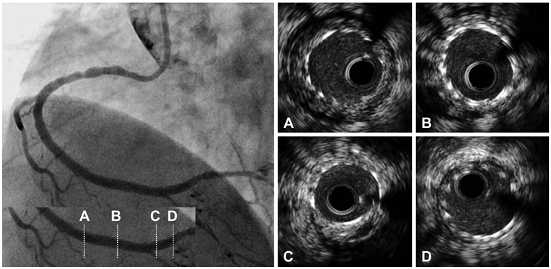
Fig. 3 Final coronary angiogram demonstrated no residual lumen narrowing with thrombolysis in myocardial infarction 3 flow. Post-stenting intravascular ultrasound images revealed well-opposed stent struts to the vessel wall (A-D) and a 4.66 mm2 minimal stent area (C).
Reference
-
1. Maehara A, Mintz GS, Bui AB, et al. Incidence, morphology, angiographic findings, and outcomes of intramural hematomas after percutaneous coronary interventions: an intravascular ultrasound study. Circulation. 2002. 105:2037–2042.2. Mahr P, Ge J, Haude M, Gorge G, Erbel R. Extramural vessel wall hematoma causing a reduced vessel diameter after coronary stenting: diagnosis by intravascular ultrasound and treatment by stent implantation. Cathet Cardiovasc Diagn. 1998. 43:438–443.3. Hirose M, Kobayashi Y, Kreps EM, et al. Luminal narrowing due to intramural hematoma shift from left anterior descending coronary artery to left circumflex artery. Catheter Cardiovasc Interv. 2004. 62:461–465.4. Noh HJ, Choi JH, Song YB, et al. Intravascular ultrasound-guided troubleshooting in a large hematoma treated with fenestration using a cutting balloon. Korean Circ J. 2009. 39:171–174.5. Oesterle SN, Limpijankit T, Yeung AC, et al. Ultrasound logic: the value of intracoronary imaging for the interventionist. Catheter Cardiovasc Interv. 1999. 47:475–490.6. Mintz GS, Nissen SE, Anderson WD, et al. American College of Cardiology Clinical Expert Consensus Document on Standards for Acquisition, Measurement and Reporting of Intravascular Ultrasound Studies (IVUS): a report of the American College of Cardiology Task Force on Clinical Expert Consensus Documents. J Am Coll Cardiol. 2001. 37:1478–1492.7. Fujii K, Ochiai M, Mintz GS, et al. Procedural implications of intravascular ultrasound morphologic features of chronic total coronary occlusions. Am J Cardiol. 2006. 97:1455–1462.8. Egglin TK, Dickey KW, Rosenblatt M, Pollak JS. Retrieval of intravascular foreign bodies: experience in 32 cases. AJR Am J Roentgenol. 1995. 164:1259–1264.9. Goldberg SL, Colombo A, Maiello L, Borrione M, Finci L, Almagor Y. Intracoronary stent insertion after balloon angioplasty of chronic total occlusions. J Am Coll Cardiol. 1995. 26:713–719.10. Murphy DA, Craver JM, King SB 3rd. Distal coronary artery dissection following percutaneous transluminal coronary angioplasty. Ann Thorac Surg. 1984. 37:473–478.11. Sanchez-Recalde A, Moreno R, Jimenez-Valero S. Stenting of spontaneous intramural coronary haematoma: long-term consequences. Eur Heart J. 2008. 29:1593.12. Souteyrand G, Motreff P. Acute vessel occlusion after coronary stenting: intravascular ultrasound diagnosis of extensive intramural haematoma. Eur Heart J. 2008. 29:9.13. Werner GS, Figulla HR, Grosse W, Kreuzer H. Extensive intramural hematoma as the cause of failed coronary angioplasty: diagnosis by intravascular ultrasound and treatment by stent implantation. Cathet Cardiovasc Diagn. 1995. 36:173–178.
- Full Text Links
-
- Actions
-
Cited
- CITED
-
- Close
- Share
-
- Similar articles
-
- Sixteen years progress in recanalization of chronic carotid artery occlusion: A comprehensive review
- Late Spontaneous Recanalization of Chronic Middle Cerebral Artery Occlusion
- Evolution of Endovascular Therapy in Acute Stroke: Implications of Device Development
- Effect of Intraarterially Administered Abciximab(Glycoprotein IIb-IIIa Inhibitor) in the Acute Cerebral Vascular Occlusion
- Recanalization of Portal Vein Graft Occlusion via a Percutaneous Transmesenteric Approach: A Case Report
