J Korean Surg Soc.
2012 Aug;83(2):111-114. 10.4174/jkss.2012.83.2.111.
Primary gastric Hodgkin's lymphoma
- Affiliations
-
- 1Department of Hospital Pathology, The Catholic University of Korea St. Vincent Hospital, Suwon, Korea.
- 2Department of Surgery, The Catholic University of Korea St. Vincent Hospital, Suwon, Korea. hchin@catholic.ac.kr
- KMID: 1820097
- DOI: http://doi.org/10.4174/jkss.2012.83.2.111
Abstract
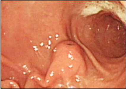
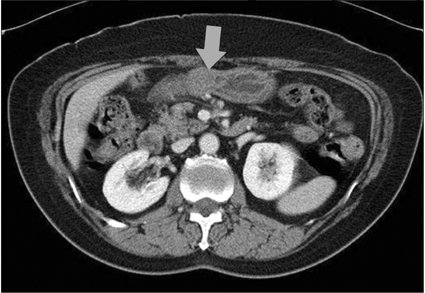
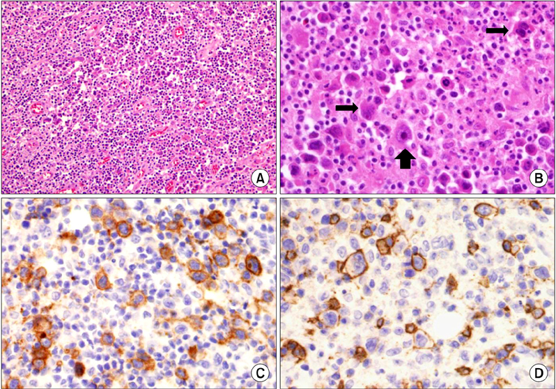
- Gastric Hodgkin's lymphoma is extremely rare. We present a case of primary Hodgkin's lymphoma arising in the stomach of a 65-year-old woman. The patient complained of epigastric discomfort and reflux for one month. Endoscopic examination revealed a protruding lesion characterized by a smooth surface at the antrum. An abdominal computed tomography uncovered a 2.5 x 2.0 cm, exophytic submucosal mass. After the tentative preoperative diagnosis of a gastrointestinal stromal tumor, a gastric wedge resection was performed. Microscopic examination of the mass demonstrated a diffuse proliferation of large atypical lymphoid cells with mono- and binucleated pleomorphic nuclei and prominent nucleoli. Immunohistochemically, the tumor cells were positive for CD30, CD20, and CD79a, whereas they were negative for cytokeratin, carcinoembryonic antigen, CD3, CD15, epithelial membrane antigen, and anaplastic lymphoma kinase-1. Based on the morphological features and immunohistochemical results, in addition to the clinical findings, a diagnosis of primary gastric Hodgkin's lymphoma was established.
Keyword
MeSH Terms
Figure
Reference
-
1. Min HR, Shin YM, Lee SD, Chun BK. Clinical study of primary gastric lymphoma and analysis of prognostic factors. J Korean Surg Soc. 1999. 56:906–914.2. Colucci G, Giotta F, Maiello E, Fucci L, Caruso ML. Primary Hodgkin's disease of the stomach: a case report. Tumori. 1992. 78:280–282.3. Devaney K, Jaffe ES. The surgical pathology of gastrointestinal Hodgkin's disease. Am J Clin Pathol. 1991. 95:794–801.4. Ogawa Y, Chung YS, Nakata B, Muguruma K, Fujimoto Y, Yoshikawa K, et al. A case of primary Hodgkin's disease of the stomach. J Gastroenterol. 1995. 30:103–107.5. Mori N, Yatabe Y, Narita M, Hayakawa S, Ishido T, Kikuchi M, et al. Primary gastric Hodgkin's disease. Morphologic, immunohistochemical, and immunogenetic analyses. Arch Pathol Lab Med. 1995. 119:163–166.6. Doweiko J, Dezube BJ, Pantanowitz L. Unusual sites of Hodgkins lymphoma: CASE 1. HIV-associated Hodgkin's lymphoma of the stomach. J Clin Oncol. 2004. 22:4227–4228.7. Venizelos I, Tamiolakis D, Bolioti S, Nikolaidou S, Lambropoulou M, Alexiadis G, et al. Primary gastric Hodgkin's lymphoma: a case report and review of the literature. Leuk Lymphoma. 2005. 46:147–150.8. Hossain FS, Koak Y, Khan FH. Primary gastric Hodgkin's lymphoma. World J Surg Oncol. 2007. 5:119.9. Saito M, Tanaka S, Mori A, Toyoshima N, Irie T, Morioka M. Primary gastric Hodgkin's lymphoma expressing a B-Cell profile including Oct-2 and Bob-1 proteins. Int J Hematol. 2007. 85:421–425.10. Dawson IM, Cornes JS, Morson BC. Primary malignant lymphoid tumours of the intestinal tract. Report of 37 cases with a study of factors influencing prognosis. Br J Surg. 1961. 49:80–89.
- Full Text Links
- Actions
-
Cited
- CITED
-
- Close
- Share
- Similar articles
-
- Case of Synchronous Primary Gastric Diffuse Large B-Cell Lymphoma and Hepatocellular Carcinoma
- Primary Non-Hodgkin T-Cell Lymphoma of the Esophagus
- A Case of Primary Non-Hodgkin's Lymphoma of the Ovary
- Rare Case of Primary Gastric Burkitt Lymphoma in a Child
- A Case of Primary Gastric Lymphoma in Puberty


