Spontaneous Regression of Focal Nodular Hyperplasia of the Liver after Delivery: A Case Report
- Affiliations
-
- 1Department of Radiology, Soonchunhyang University Bucheon Hospital, Soonchunhyang University College of Medicine, Bucheon, Korea. radmhlee@schmc.ac.kr
- 2Department of Pathology, Soonchunhyang University Bucheon Hospital, Soonchunhyang University College of Medicine, Bucheon, Korea.
- KMID: 1819781
- DOI: http://doi.org/10.3348/jksr.2014.71.2.65
Abstract
- A focal nodular hyperplasia (FNH) of the liver is the second most common benign hepatic tumor, but an interval growth or regression of FNH is uncommon. We report a case of spontaneous regression of FNH in a 34-year-old woman. This lesion was diagnosed during pregnancy and spontaneously regressed during a 5-year observation period after delivery.
Figure
-
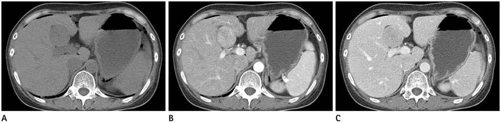
Fig. 1 A dynamic abdominal CT scan acquired five days after the delivery A. Precontrast CT scan shows a 4.2 × 4.0 cm size, well defined exophytic growing low density mass arising from segment IV of liver. B, C. On arterial phase, the mass is well enhanced (B), and shows slightly low attenuation compared with the surrounding liver parenchyma on portal phase (C).
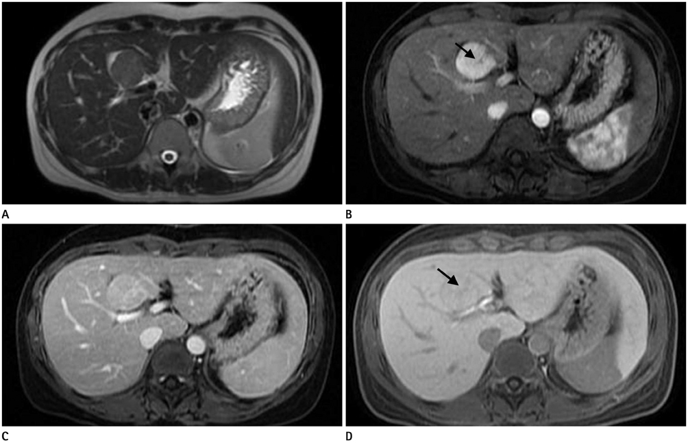
Fig. 2 Liver MRI using hepatocyte-specific contrast agent. A. A T2-weighted imaging shows the high SI mass at segment IV. B. Following intravenous administration of gadobenate dimeglumine, an arterial phase T1-weighted imaging (T1WI) shows an intensely enhancing mass with a nonenhancing central scar (arrow). C. Portal phase image shows that the mass is isointense with the liver parenchyma and the central scar enhancing. D. A hepatobiliary phase T1WI shows an isointense mass with a hypointense central scar (arrow).
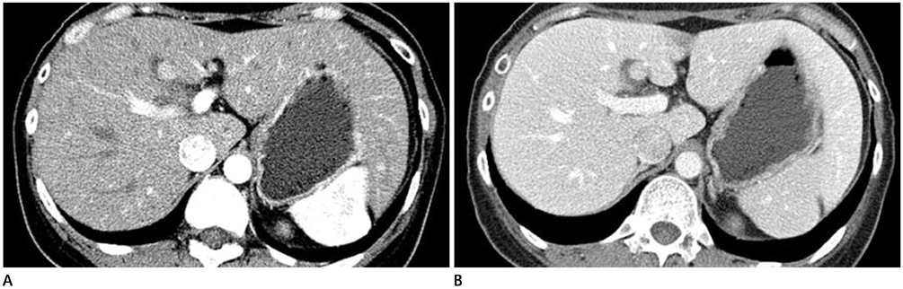
Fig. 3 On follow-up CT scan acquired 5 years later, the focal nodular hyperplasia is measured 0.8 × 1.0 cm. This lesion still shows intense enhancement on the arterial phase (A) and iso-density on the portal phase (B).
Reference
-
1. Wanless IR, Mawdsley C, Adams R. On the pathogenesis of focal nodular hyperplasia of the liver. Hepatology. 1985; 5:1194–1200.2. Buetow PC, Pantongrag-Brown L, Buck JL, Ros PR, Goodman ZD. Focal nodular hyperplasia of the liver: radiologic-pathologic correlation. Radiographics. 1996; 16:369–388.3. Côté C. Regression of focal nodular hyperplasia of the liver after oral contraceptive discontinuation. Clin Nucl Med. 1997; 22:587–590.4. Di Stasi M, Caturelli E, De Sio I, Salmi A, Buscarini E, Buscarini L. Natural history of focal nodular hyperplasia of the liver: an ultrasound study. J Clin Ultrasound. 1996; 24:345–350.5. Mathieu D, Kobeiter H, Maison P, Rahmouni A, Cherqui D, Zafrani ES, et al. Oral contraceptive use and focal nodular hyperplasia of the liver. Gastroenterology. 2000; 118:560–564.6. Leconte I, Van Beers BE, Lacrosse M, Sempoux C, Jamart J, Materne R, et al. Focal nodular hyperplasia: natural course observed with CT and MRI. J Comput Assist Tomogr. 2000; 24:61–66.7. Giannitrapani L, Soresi M, La Spada E, Cervello M, D'Alessandro N, Montalto G. Sex hormones and risk of liver tumor. Ann N Y Acad Sci. 2006; 1089:228–236.8. Cobey FC, Salem RR. A review of liver masses in pregnancy and a proposed algorithm for their diagnosis and management. Am J Surg. 2004; 187:181–191.9. Rifai K, Mix H, Krusche S, Potthoff A, Manns MP, Gebel MJ. No evidence of substantial growth progression or complications of large focal nodular hyperplasia during pregnancy. Scand J Gastroenterol. 2013; 48:88–92.10. Halankar JA, Kim TK, Jang HJ, Khalili K, Masoom HA. Understanding the natural history of focal nodular hyperplasia in the liver with MRI. Indian J Radiol Imaging. 2012; 22:116–120.
