Subarachnoid Hemorrhage with Negative Baseline Digital Subtraction Angiography: Is Repeat Digital Subtraction Angiography Necessary?
- Affiliations
-
- 1Department of Neurosurgery, College of Medicine, Yeungnam University, Daegu, Korea. cch0102@ynu.ac.kr
- KMID: 1808457
- DOI: http://doi.org/10.7461/jcen.2012.14.3.210
Abstract
- INTRODUCTION
Patients with negative initial digital subtraction angiography (DSA) are at significant risk for re-bleeding, which can lead to severe disability and death. The purpose of this study was to evaluate the necessity of repeat DSA in subgroups of patients with subarachnoid hemorrhage (SAH) with negative initial DSA.
METHODS
A total of 904 spontaneous SAH patients were admitted to our department between May 2005 and May 2012. Twenty eight patients were selected for inclusion in this study because repeated DSA performed due to the etiology of the SAH could not be demonstrated on the initial DSA. According to the SAH pattern on initial computed tomography scans, patients were divided into perimesencephalic nonaneurysmal SAH (PN-SAH) and non PN-SAH (NPN-SAH) groups. Repeat DSA was performed in all patients, and two of these patients underwent a third DSA.
RESULTS
Of the 904 patients, 28 patients (3.1%) had no vascular abnormality on initial DSA. Sixteen PN-SAH patients underwent a repeat DSA; however, no aneurysms were found. In contrast, 12 patients with NPN-SAH underwent repeat DSA, with detection of two cerebral aneurysms. Overall, the false-negative rate of the initial DSA was 7.1% (2/28 patients). No significant differences in false-negative results on initial DSA were observed between the PN-SAH and NPN-SAH groups.
CONCLUSION
In the line with the results of the current study, we should be highly suspicious of patients with a nonaneurysmal SAH, especially those with a NPN-SAH pattern. In order to reduce the morbidity and mortality resulting from a misdiagnosis, repeat DSA is necessary, and exclusion of an aneurysm is important.
MeSH Terms
Figure
-
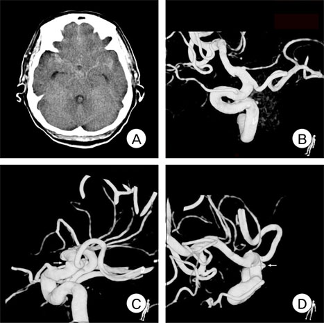
Fig. 1 Initial noncontrast brain computed tomography (CT) and digital subtraction angiography (DSA). Diffuse subarachnoid hemorrhage (SAH) in the region of both the sylvian fissure and interhemispheric fissure (A). Initial three-dimensional rotational angiography image reveals no aneurysmal structure (B). Repeat DSA was performed on the eighth day of admission. Three-dimensional rotational angiography images clearly show a distal ICA aneurysm (C, D).
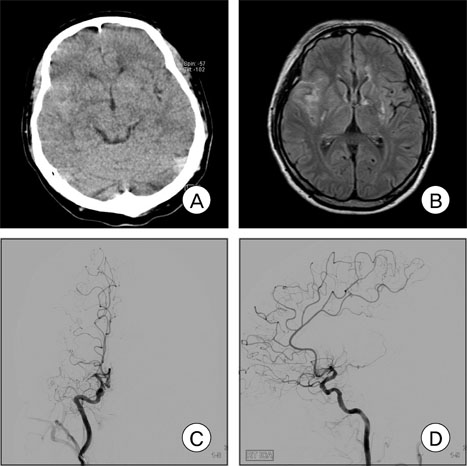
Fig. 2 Initial brain CT, magnetic resonance imaging (MRI) and DSA on admission. Brain noncontrast CT and MRI flair images show a subarachnoid hemorrhage on the right sylvian fissure (A, B), Mid-arterial phase DSA images show occlusion of the right M1 segment of the middle cerebral artery, but reveal no aneurysmal structure (C, D).
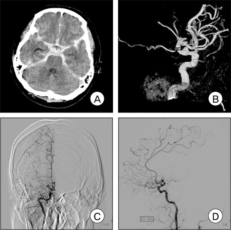
Fig. 3 Brain CT and repeat DSA on the sixth day of admission. A large amount of SAH is observed in the region of the basal cistern, both sylvian fissures, and interhemispheric fissure (A). Repeat DSA and three-dimensional angiography show an aneurysm on the ophthalmic segment of the right internal cerebral artery, probably a blister-like dorsal wall aneurysm (B, C, D).
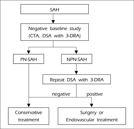
Fig. 4 Flow-sheet for diagnosis of patients with subarachnoid hemorrhage.
Cited by 1 articles
-
Arterial Wall Imaging in Angiographically Occult Spontaneous Subarachnoid Hemorrhage : New Insight into the Usual Suspect
Wonki Yoon, Jang Hun Kim, Haewon Roh, Taek-Hyun Kwon
J Korean Neurosurg Soc. 2022;65(2):245-254. doi: 10.3340/jkns.2021.0120.
Reference
-
1. Abe M, Tabuchi K, Yokoyama H, Uchino A. Blood blisterlike aneurysms of the internal carotid artery. J Neurosurg. 1998. 09. 89(3):419–424.
Article2. Broderick JP, Brott TG, Duldner JE, Tomsick T, Leach A. Initial and recurrent bleeding are the major causes of death following subarachnoid hemorrhage. Stroke. 1994. 07. 25(7):1342–1347.
Article3. Cruz JP, Sarma D, Noel de Tilly L. Perimesencephalic subarachnoid hemorrhage: when to stop imaging? Emerg Radiol. 2011. 06. 18(3):197–202.
Article4. Ishihara H, Kato S, Akimura T, Suehiro E, Oku T, Suzuki M. Angiogram-negative subarachnoid hemorrhage in the era of three dimensional rotational angiography. J Clin Neurosci. 2007. 03. 14(3):252–255.
Article5. Kaufmann TJ, Huston J 3rd, Mandrekar JN, Schleck CD, Thielen KR, Kallmes DF. Complications of diagnostic cerebral angiography: evaluation of 19,826 consecutive patients. Radiology. 2007. 06. 243(3):812–819.6. McMahon J, Dorsch N. Subarachnoid haemorrhage of unknown aetiology: what next? Crit Rev Neurosurg. 1999. 05. 9(3):147–155.
Article7. Ogawa A, Suzuki M, Ogasawara K. Aneurysms at nonbranching sites in the supraclinoid portion of the internal carotid artery: internal carotid artery trunk aneurysms. Neurosurgery. 2000. 09. 47(3):578–583. discussion 83-6.
Article8. Rinkel GJ, Wijdicks EF, Vermeulen M, Hageman LM, Tans JT, van Gijn J. Outcome in perimesencephalic (nonaneurysmal) supraclinoid hemorrhage: a follow-up study in 37 patients. Neurology. 1990. 07. 40(7):1130–1132.9. Rinkel GJ, Wijdicks EF, Hasan D, Kienstra GE, Franke CL, Hageman LM, et al. Outcome in patients with subarachnoid haemorrhage and negative angiography according to pattern of haemorrhage on computed tomography. Lancet. 1991. 10. 338(8773):964–968.
Article10. Rinkel GJ, Wijdicks EF, Vermeulen M, Ramos LM, Tanghe HL, Hasan D, et al. Nonaneurysmal perimesencephalic subarachnoid hemorrhage: CT and MR patterns that differ from aneurysmal rupture. AJNR Am J Neuroradiol. 1991. Sep-Oct. 12(5):829–834.11. Topcuoglu MA, Ogilvy CS, Carter BS, Buonanno FS, Koroshetz WJ, Singhal AB. Subarachnoid hemorrhage without evident cause on initial angiography studies: diagnostic yield of subsequent angiography and other neuroimaging tests. J Neurosurg. 2003. 06. 98(6):1235–1240.
Article12. van Gijn J, Rinkel GJ. Subarachnoid haemorrhage: diagnosis, causes and management. Brain. 2001. 02. 124(Pt 2):249–278.
Article13. van Gijn J, van Dongen KJ, Vermeulen M, Hijdra A. Perimesencephalic hemorrhage: a nonaneurysmal and benign form of subarachnoid hemorrhage. Neurology. 1985. 04. 35(4):493–497.
Article14. Velthuis BK, Rinkel GJ, Ramos LM, Witkamp TD, van Leeuwen MS. Perimesencephalic hemorrhage. Exclusion of vertebrobasilar aneurysms with CT angiography. Stroke. 1999. 05. 30(5):1103–1109.
- Full Text Links
-
- Actions
-
Cited
- CITED
-
- Close
- Share
-
- Similar articles
-
- Cerebral Venous Thrombosis Presenting as Isolated Subarachnoid Hemorrhage
- A Clinical Significance of lntraarterial Digital Subtraction Angiography in Renal Diseases
- Clinical Applications of Digital Subtraction Angiography(DSA) in Neurosurgical Field
- Radiologic consideration of intra-arterial digital subtraction angiography
- Rupture of De Novo Anterior Communicating Artery Aneurysm 8 Days after the Clipping of Ruptured Middle Cerebral Artery Aneurysm
