J Cerebrovasc Endovasc Neurosurg.
2012 Sep;14(3):170-174. 10.7461/jcen.2012.14.3.170.
Stereotactic Burr Hole Aspiration Surgery for Spontaneous Hypertensive Cerebellar Hemorrhage
- Affiliations
-
- 1Department of Neurosurgery, Institute of Wonkwang Medical Science, School of Medicine, Wonkwang University, Iksan, Korea. kangsd@wku.ac.kr
- KMID: 1808451
- DOI: http://doi.org/10.7461/jcen.2012.14.3.170
Abstract
OBJECTIVE
Patients with severe spontaneous cerebellar hemorrhage typically undergo treatment with suboccipital craniectomy and hematoma evacuation. However, this is a stressful procedure for patients due to the long operating time and operation-induced tissue damage. In addition, the durotomy can result in pseudomeningocele. We investigated the efficacy of stereotactic or navigation-guided burr hole aspiration surgery as a treatment for spontaneous hypertensive cerebellar hemorrhage (SHCH).
METHODS
Between January 2002 and December 2011, 26 patients with SHCH underwent surgery using the stereotactic or navigation-guided burr hole aspiration and catheter insertion technique in our institution.
RESULTS
Mean hematoma volume was 21.8 +/- 5.8 cc at admission and 13.1 +/- 5.4 cc immediately following surgery. Preoperative Glasgow Coma Scale (GCS) score was 12.5 +/- 1.3 and postoperative GCS score was 13.1 +/- 1.2. Seven days after surgery, the mean hematoma volume was 4.3 +/- 5.6 cc, and there was no occurrence of surgery-related complications during the six-month follow-up period. The mean operation time for catheter insertion was 43.1 +/- 8.9 min, and a mean 31.3 +/- 6.0 min was also added for extra-ventricular drainage. The mean Glasgow Outcome Scale (GOS) score after six months was 4.6 +/- 1.0.
CONCLUSION
Stereotactic burr hole aspiration surgery for treatment of SHCH is less time-consuming and invasive than other interventions, and resulted in no surgery-related complications. Therefore, we suggest that this surgical method could be a safe and effective treatment option for selected patients with SHCH.
Keyword
MeSH Terms
Figure
-
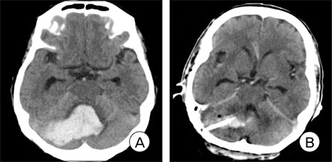
Fig. 1 A 56-year-old male patient presented with a drowsy mental status. (A) Findings on brain Computed tomography (CT) at admission shows spontaneous cerebellar hemorrhage with mild brain stem compression and obstruction of the fourth ventricle. The hematoma volume was 26 cc and maximal hematoma diameter was 49 mm. (B) Brain CT at three days after surgery shows residual hematoma with catheter after stereotactic aspiration surgery.
Reference
-
1. Brennan RW, Bergland RM. Acute cerebellar hemorrhage. Analysis of clinical findings and outcome in 12 cases. Neurology. 1977. 06. 27(6):527–532.
Article2. Broderick JP, Adams HP Jr, Barsan W, Feinberg W, Feldmann E, Grotta J, et al. Guidelines for the management of spontaneous intracerebral hemorrhage: A statement for healthcare professionals from a special writing group of the Stroke Council, American Heart Association. Stroke. 1999. 04. 30(4):905–915.3. Broderick J, Connolly S, Feldmann E, Hanley D, Kase C, Krieger D, et al. Guidelines for the management of spontaneous intracerebral hemorrhage in adults: 2007 update: a guideline from the American Heart Association/American Stroke Association Stroke Council, High Blood Pressure Research Council, and the Quality of Care and Outcomes in Research Interdisciplinary Working Group. Circulation. 2007. 10. 116(16):e391–e413.4. Chin D, Carney P. Acute cerebellar hemorrhage with brainstem compression in contrast with benign cerebellar hemorrhage. Surg Neurol. 1983. 05. 19(5):406–409.
Article5. Donauer E, Loew F, Faubert C, Alesch F, Schaan M. Prognostic factors in the treatment of cerebellar hemorrhage. Acta Neurochir (Wien). 1994. 131(1-2):59–66.6. Firsching R, Huber M, Frowein RA. Cerebellar hemorrhage: Management and prognosis. Neurosurg Rev. 1991. 14(3):191–194.7. Heros RC. Cerebellar hemorrhage and infarction. Stroke. 1982. Jan-Feb. 13(1):106–109.
Article8. Kobayashi S, Sato A, Kageyama Y, Nakamura H, Watanabe Y, Yamaura A. Treatment of hypertensive cerebellar hemorrhage-Surgical or conservative management? Neurosurgery. 1994. 02. 34(2):246–250. discussion 250-1.
Article9. Koziarski A, Frankiewicz E. Medical and surgical treatment of intracerebellar hematomas. Acta Neurochir (Wien). 1991. 110(1-2):24–28.10. Lui TN, Fairholm DJ, Shu TF, Chang CN, Lee ST, Chen HR. Surgical treatment of spontaneous cerebellar hemorrhage. Surg Neurol. 1985. 06. 23(6):555–558.
Article11. Mezzadri JJ, Otero JM, Ottino CA. Management of 50 spontaneous cerebellar hemorrhages. Importance of obstructive hydrocephalus. Acta Neurochir (Wien). 1993. 122(1-2):39–44.12. Mohadjer M, Eggert R, May J, Mayfrank L. CT-guided stereotactic fibrinolysis of spontaneous and hypertensive cerebellar hemorrhage: long term results. J Neurosurg. 1990. 08. 73(2):217–222.13. Niizuma H, Suzuki J. Computed tomography-guided stereotactic aspiration of posterior fossa hematomas: a supine lateral retromastoid approach. Neurosurgery. 1987. 09. 21(3):422–427.
Article14. Norris JW, Eisen AA, Branch CL. Problems in cerebellar hemorrhage and infarction. Neurology. 1969. 11. 19(11):1043–1050.
Article15. Salazar J, Vaquero J, Martinez P, Santos H, Martinez R, Bravo G. Clinical and CT scan assessment of benign versus fatal spontaneous cerebellar hematomas. Acta Neurochir (Wien). 1986. 79(2-4):80–86.16. Taneda M, Hayakawa T, Mogami H. Primary cerebellar hemorrhage. Quadrigeminal cistern obliteration on CT scans as a predictor of outcome. J Neurosurg. 1987. 10. 67(4):545–552.17. Winn HR. Youmans Neurological Surgery. 2011. ed 6. Philadelphia: W.B. Saunders Co;3712.18. Yanaka K, Meguro K, Fujita K, Narushima K, Nose T. Immediate surgery reduces mortality in deeply comatose patients with spontaneous cerebellar hemorrhage. Neurol Med Chir (Tokyo). 2000. 06. 40(6):295–299. discussion 299-300.
- Full Text Links
-
- Actions
-
Cited
- CITED
-
- Close
- Share
-
- Similar articles
-
- Cerebellar Hemorrhage after Burr Hole Drainage of Supratentorial Chronic Subdural Hematoma
- Delayed Cerebellar Hemorrhage after Supratentorial Burr-Hole Drainage
- Stereotactic Management of Spontaneous Infratenorial Hemorrhage: Part II: Transtentorial Stereotactic Approach for Spontaneous Intracerebellar Hemorrhage
- Safe and time-saving treatment method for acute cerebellar infarction: Navigation-guided burr-hole aspiration – 6-years single center experience
- Risk Factors in Patients with Rebleeding after CT-Guided Stereotactic Burr Hole Aspiration for Hypertensive Intracerebral Hematoma
