Predominant Hypertensive Brainstem Encephalopathy with Supratentorial Involvement: Case Report and Literature Review
- Affiliations
-
- 1Department of Radiology, Soonchunhyang University Hospital, Soonchunhyang University School of Medicine, Seoul, Korea. stpark@schmc.ac.kr
- 2Department of Radiology, Samsung Medical Center, Sungkyunkwan University School of Medicine, Seoul, Korea.
- KMID: 1801555
- DOI: http://doi.org/10.3348/jksr.2014.71.6.314
Abstract
- Hypertensive encephalopathy typically presents with bilateral parietooccipital vasogenic edema. Brainstem and cerebellar edema are uncommon in association with typical supratentorial changes. We experienced three cases of atypical hypertensive encephalopathy involving brainstem and cerebellum as well as cerebral white matter, which showed characteristic alternating linear bright and low signals in the pons, the so-called "stripe sign". We report these cases here with a brief literature review.
MeSH Terms
Figure
-

Fig. 1 A 36-year-old female presented with headache. MR images showed hyperintensity involving the periventricular cerebral white matter (A), pons and cerebellum bilaterally, with the stripe sign in the pons on fluid-attenuated inversion recovery (FLAIR) images (B). Corresponding apparent diffusion coefficient map revealed increased diffusion in the brainstem (C). Follow-up MR images 3 weeks after initial MRI showed significant diminution of hyperintensity on FLAIR images.
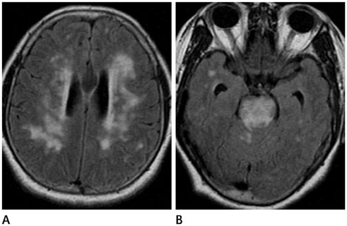
Fig. 2 A 55-year-old female suffered from headache and vomiting. MR images showed symmetric hyperintensity in the periventricular cerebral white matter (A), brainstem and cerebellum on fluid-attenuated inversion recovery images with the stripe sign in the pons. The periphery of the pons was relatively spared (B).
Reference
-
1. Bhagavati S, Chum F, Choi J. Hypertensive encephalopathy presenting with isolated brain stem and cerebellar edema. J Neuroimaging. 2008; 18:454–456.2. Ono Y, Manabe Y, Hamakawa Y, Murakami T, Omori N, Hayashi Y, et al. Localized lesions on MRI in a case of hypertensive brainstem encephalopathy. Intern Med. 2005; 44:1002–1005.3. de Seze J, Mastain B, Stojkovic T, Ferriby D, Pruvo JP, Destée A, et al. Unusual MR findings of the brain stem in arterial hypertension. AJNR Am J Neuroradiol. 2000; 21:391–394.4. Osborn AG. Acquired Metabolic and Systemic Disorders. In : Osborn AG, editor. Osborn's Brain: Imaging, Pathology, and Anatomy. Salt Lake City, UT: Amirsys;2012. p. 907–957.5. Beausang-Linder M, Bill A. Cerebral circulation in acute arterial hypertension--protective effects of sympathetic nervous activity. Acta Physiol Scand. 1981; 111:193–199.6. Sadoshima S, Fujii K, Yao H, Kusuda K, Ibayashi S, Fujishima M. Regional cerebral blood flow autoregulation in normotensive and spontaneously hypertensive rats--effects of sympathetic denervation. Stroke. 1986; 17:981–984.7. Karasawa S, Kawanami T, Kimura H, Kurita K, Kato T. An unusual case of hypertensive encephalopathy involving the brain stem. Intern Med. 2004; 43:448–449.8. Kanazawa M, Sanpei K, Kasuga K. Recurrent hypertensive brainstem encephalopathy. J Neurol Neurosurg Psychiatry. 2005; 76:888–890.9. Gamanagatti S, Subramanian S. Hypertensive encephalopathy: isolated pons involvement mimicking central pontine myelinolysis. Korean J Radiol. 2006; 7:218–219.10. Uchino M, Haga D, Nomoto J, Mito T, Kuramitsu T. Brainstem involvement in hypertensive encephalopathy: a report of two cases and literature review. Eur Neurol. 2007; 57:223–226.11. Kang SY, Choi JC, Kang JH. Two cases of hypertensive encephalopathy involving the brainstem. J Clin Neurol. 2007; 3:50–52.12. Shintani S, Hino T, Ishihara S, Mizutani S, Shiigai T. Reversible brainstem hypertensive encephalopathy (RBHE): clinicoradiologic dissociation. Clin Neurol Neurosurg. 2008; 110:1047–1053.13. Chang GY, Keane JR. Hypertensive brainstem encephalopathy: three cases presenting with severe brainstem edema. Neurology. 1999; 53:652–654.14. Yasuda Y, Akiguchi I, Imai T, Sonobe M, Kage M. Hypertensive brainstem encephalopathy. Intern Med. 2003; 42:1131–1134.15. Nagata M, Maeda M, Tsukahara H, Maier SE, Takeda K. Brain stem hypertensive encephalopathy evaluated by line scan diffusion-weighted imaging. AJNR Am J Neuroradiol. 2004; 25:803–806.16. Doi Y, Kimura F, Fujiyama T, Fujimura C, Nishina T, Sato T, et al. Hypertensive brainstem encephalopathy without parieto-occipital lesion--two case reports. Neurol Med Chir (Tokyo). 2006; 46:75–79.17. Yoshida K, Yamamoto T, Mori K, Maeda M. Reversible posterior leukoencephalopathy syndrome in a patient with hypertensive encephalopathy--case report. Neurol Med Chir (Tokyo). 2001; 41:364–369.18. Kumai Y, Toyoda K, Fujii K, Ibayashi S. Hypertensive encephalopathy extending into the whole brainstem and deep structures. Hypertens Res. 2002; 25:797–800.19. Cruz-Flores S, de Assis Aquino Gondim F, Leira EC. Brainstem involvement in hypertensive encephalopathy: clinical and radiological findings. Neurology. 2004; 62:1417–1419.20. Park JH, Kim SM, Shin HW, An SJ. Hypertensive brainstem encephalopathy involving deep supratentorial regions: does only blood pressure matter? Neurol Int. 2010; 2:e9.
- Full Text Links
-
- Actions
-
Cited
- CITED
-
- Close
- Share
-
- Similar articles
-
- Two Cases of Hypertensive Encephalopathy Involving the Brainstem
- Hypertensive Brainstem Encephalopathy with Atypical Supratentorial Involvement
- Hypertensive Brainstem Encephalopathy with Extensive Supratentorial Involvement
- Two Cases of Hypertensive Brainstem Encephalopathy
- Reversible Obstructive Hydrocephalus Associated with Hypertensive Brainstem Encephalopathy
