Eosinophilic Gastroenteritis Due to Rhus Ingestion Presenting with Gastrointestinal Hemorrhage
- Affiliations
-
- 1Department of Internal Medicine, Chonnam National University Medical School, Gwangju, Korea. drpsy@naver.com
- 2Department of Pathology, Chonnam National University Medical School, Gwangju, Korea.
- KMID: 1801211
- DOI: http://doi.org/10.5946/ce.2015.48.2.174
Abstract
- Rhus-related illnesses in Korea are mostly caused by ingestion of parts of the Rhus tree. Contact dermatitis occurrence after ingestion of Rhus-related food is very common in Korea. However, Rhus-related gastrointestinal disease is very rare. Herein, we present a case of eosinophilic gastroenteritis caused by Rhus ingestion. A 75-year-old woman was admitted with hematemesis and hematochezia after Rhus extract ingestion. Routine laboratory tests revealed leukocytosis without eosinophilia. Endoscopy showed friable and granular mucosal changes with touch bleeding in the second portion of the duodenum. Abdominal computed tomography revealed edematous wall thickening of the duodenum and proximal jejunal loops. Patch testing with Rhus extracts showed a strong positive reaction, suggesting Rhus as the allergen. Her symptoms improved after avoidance of the allergen.
MeSH Terms
Figure
-
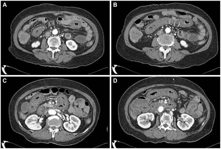
Fig. 1 (A-D) Abdominal computed tomography image showing edematous wall thickening in the duodenum and proximal jejunal loops.
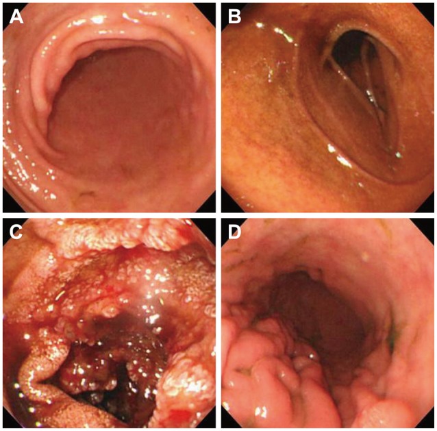
Fig. 2 (A-D) Endoscopy image showing friable and granular mucosal changes with touch bleeding in the second portion of the duodenum. However, there was no involvement of the stomach and duodenal bulb.
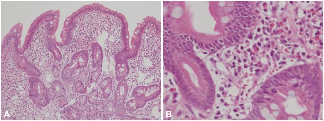
Fig. 3 Pathologic findings showing edematous gastric mucosa and diffusely infiltrated inflammatory cells containing over 100 eosinophils per high power field, consistent with eosinophilic gastroenteritis (A: H&E stain, ×100; B: H&E stain, ×400).
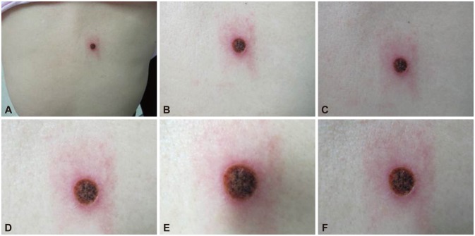
Fig. 4 (A-F) Patch test. A Rhus extract-containing patch was applied for 48 hours, and then removed. Seventy-two hours after application, the skin showed erythematous infiltration with ulceration, which is a strongly positive reaction.
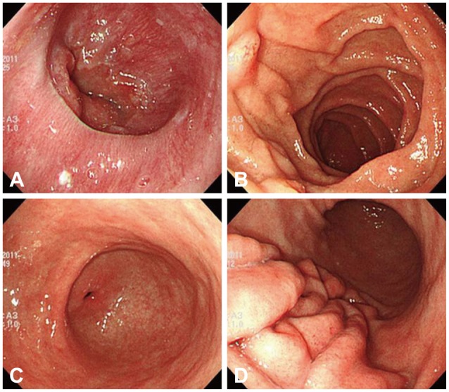
Fig. 5 (A-D) Follow-up endoscopy image showing continued improvement of the mucosal edema and nodularity in the duodenum.
Reference
-
1. Yun SK, Ko KB, Song IM, Choi SP, Ihm CW. Epidemiologic study on systemic contact dermatitis due to ingestion of Rhus. Korean J Dermatol. 2002; 40:253–257.2. Yoo KH, Seo SJ, Li K, Hong CK. Ingestion of Rhus chicken causing systemic contact dermatitis in a Korean patient. Clin Exp Dermatol. 2010; 35:756–758. PMID: 20456389.
Article3. Park SD, Lee SW, Chun JH, Cha SH. Clinical features of 31 patients with systemic contact dermatitis due to the ingestion of Rhus (lacquer). Br J Dermatol. 2000; 142:937–942. PMID: 10809851.
Article4. Ahn BM. Rhus-chicken. Korean J Hepatol. 2002; 8:245–247.5. Coombes JL, Powrie F. Dendritic cells in intestinal immune regulation. Nat Rev Immunol. 2008; 8:435–446. PMID: 18500229.
Article6. Khan S. Eosinophilic gastroenteritis. Best Pract Res Clin Gastroenterol. 2005; 19:177–198. PMID: 15833687.
Article7. Kinoshita Y, Furuta K, Ishimaura N, et al. Clinical characteristics of Japanese patients with eosinophilic esophagitis and eosinophilic gastroenteritis. J Gastroenterol. 2013; 48:333–339. PMID: 22847555.
Article8. Rothenberg ME, Mishra A, Brandt EB, Hogan SP. Gastrointestinal eosinophils in health and disease. Adv Immunol. 2001; 78:291–328. PMID: 11432207.
Article9. Rothenberg ME. Eosinophilic gastrointestinal disorders (EGID). J Allergy Clin Immunol. 2004; 113:11–28. PMID: 14713902.
Article10. Liacouras CA, Spergel JM, Ruchelli E, et al. Eosinophilic esophagitis: a 10-year experience in 381 children. Clin Gastroenterol Hepatol. 2005; 3:1198–1206. PMID: 16361045.
Article11. Chen MJ, Chu CH, Lin SC, Shih SC, Wang TE. Eosinophilic gastroenteritis: clinical experience with 15 patients. World J Gastroenterol. 2003; 9:2813–2816. PMID: 14669340.
Article
- Full Text Links
-
- Actions
-
Cited
- CITED
-
- Close
- Share
-
- Similar articles
-
- A Case of Eosinophilic Gastroenteritis Presenting with Peritoneal Eosinpophilic Infiltration
- A Case of Eosinophilic Gastroenteritis Associsted with Protein - losing Enteropathy
- Diffuse Eosinophilic Gastroenteritis with Antral Obstruction: A Case Report
- Serosal Type Eosinophilic Gastroenteritis
- Eosinophilic Enteritis Diagnosed by Laparoscopic Biopsy
