Ann Surg Treat Res.
2015 Jun;88(6):349-352. 10.4174/astr.2015.88.6.349.
Alternative prosthetic vascular access creation using subscapular artery as inflow to prevent dialysis access related steal syndrome
- Affiliations
-
- 1Department of Surgery, Soonchunhyang University Seoul Hospital, Soonchunhyang University College of Medicine, Seoul, Korea. ultravascsurg@gmail.com
- KMID: 1800650
- DOI: http://doi.org/10.4174/astr.2015.88.6.349
Abstract
- In patients highly suspected of developing steal syndrome, the subscapular artery may be a good supplier for functional prosthetic arteriovenous access, as well as a good solution for the prevention of steal syndrome. A 51-year-old woman was preparing to have a loop shaped polytetrafluoroethylene (PTFE) graft placed at the left upper extremity. The diameter of subscapular the artery was 3 mm. Arterial calcification was not evident. The diameter of the basilic vein was 6 mm. A 50-cm long 4-7 mm tapered PTFE graft was placed in a loop shape between both skin incisions. The patient was uneventfully discharged at postoperative day 4 without any remaining steal syndrome. The PTFE graft was well-functioning during the follow-up period. The patient did not experience symptoms of steal syndrome any longer.
Keyword
MeSH Terms
Figure
-
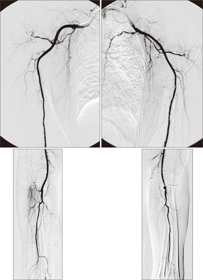
Fig. 1 Preoperative arteriogram revealed arteriopathy of upper extremity. There were only interosseous arteries without radial or ulna arteries at bilateral distal upper extremities.

Fig. 2 (A) Subscapular artery was identified and dissected from adjacent structures with good field of vision at axillary approach (black arrow). (B) Loop shaped polytetrafluoroethylene graft was placed subcutaneously between two skin incisions. (C) At 8 months postoperatively, percutaneous angioplasty was done for stenosis of graft-venous anastomosis.
Reference
-
1. Tordoir JH, Dammers R, van der Sande FM. Upper extremity ischemia and hemodialysis vascular access. Eur J Vasc Endovasc Surg. 2004; 27:1–5.2. Beathard GA, Spergel LM. Hand ischemia associated with dialysis vascular access: an individualized access flow-based approach to therapy. Semin Dial. 2013; 26:287–314.3. Song D, Moon C. Surgical treatment, with using distal revascularization intervalligation, for the ischemia that follows creation of hemodialysis access. J Korean Surg Soc. 2008; 74:371–377.4. Mickley V. Steal syndrome: strategies to preserve vascular access and extremity. Nephrol Dial Transplant. 2008; 23:19–24.5. Gradman WS, Pozrikidis C. Analysis of options for mitigating hemodialysis access-related ischemic steal phenomena. Ann Vasc Surg. 2004; 18:59–65.6. Jennings W, Brown R, Blebea J, Taubman K, Messiner R. Prevention of vascular access hand ischemia using the axillary artery as inflow. J Vasc Surg. 2013; 58:1305–1309.7. Valnicek SM, Mosher M, Hopkins JK, Rockwell WB. The subscapular arterial tree as a source of microvascular arterial grafts. Plast Reconstr Surg. 2004; 113:2001–2005.8. Bartlett SP, May JW Jr, Yaremchuk MJ. The latissimus dorsi muscle: a fresh cadaver study of the primary neurovascular pedicle. Plast Reconstr Surg. 1981; 67:631–636.9. Jennings WC, Brown RE, Ruiz C. Primary arteriovenous fistula inflow proximalization for patients at high risk for dialysis access-associated ischemic steal syndrome. J Vasc Surg. 2011; 54:554–558.10. Reifsnyder T, Arnaoutakis GJ. Arterial pressure gradient of upper extremity arteriovenous access steal syndrome: treatment implications. Vasc Endovascular Surg. 2010; 44:650–653.
- Full Text Links
-
- Actions
-
Cited
- CITED
-
- Close
- Share
-
- Similar articles
-
- Proximalization of Arterial Inflow for the Treatment of Access-Related Steal Syndrome
- Treatment of Dialysis Access Steal Syndrome with Concomitant Vascular Access Aneurysms
- Surgical Treatment, with Using Distal Revascularization Interval-Ligation, for the Ischemia that Follows Creation of Hemodialysis Access
- Successful Access Rate and Risk Factor of Vascular Access Surgery in Arm for Dialysis
- Brachial-ulnar Artery Bypass for Treating Ischemic Steal Syndrome: Report of A Case
