Intradural Extramedullary Epidermoid Cyst: A Case Report
- Affiliations
-
- 1Department of Orthopedic Surgery, Ulsan University Hospital, College of Medicine, Ulsan University, Korea. jrcha@uuh.ulsan.kr
- KMID: 1800391
- DOI: http://doi.org/10.4184/jkss.2015.22.1.26
Abstract
- STUDY DESIGN: A case report.
OBJECTIVES
To report a rare case of epidermoid cyst in the spinal canal. SUMMARY OF LITERATURE REVIEW: Epidermoid cyst in the spinal canal is rare. Idiopathic epidermoid cyst in the spinal canal not associated with a trauma or infection is even rarer. MATERIAL AND METHODS: A 73 year-old female presented with a 1 year history of progressive paresthesia and motor weakness of both lower extremeties. MRI showed a cystic mass on the 7th thoracic canal. We performed total laminectomy at the T6-T8 level. The cystic mass was excised after durotomy using a posterior approach.
RESULTS
We confirmed the presence of an epidermoid cyst for histopathology.
CONCLUSION
Idiopathic epidermoid cyst in the spine is very rare and requires accurate differential diagnosis. Preoperative MRI scans are necessary to differentiatie epidermoid cysts from other intradural masses. Confirmative diagnosis can be done by histopatholoty.
Keyword
MeSH Terms
Figure
-
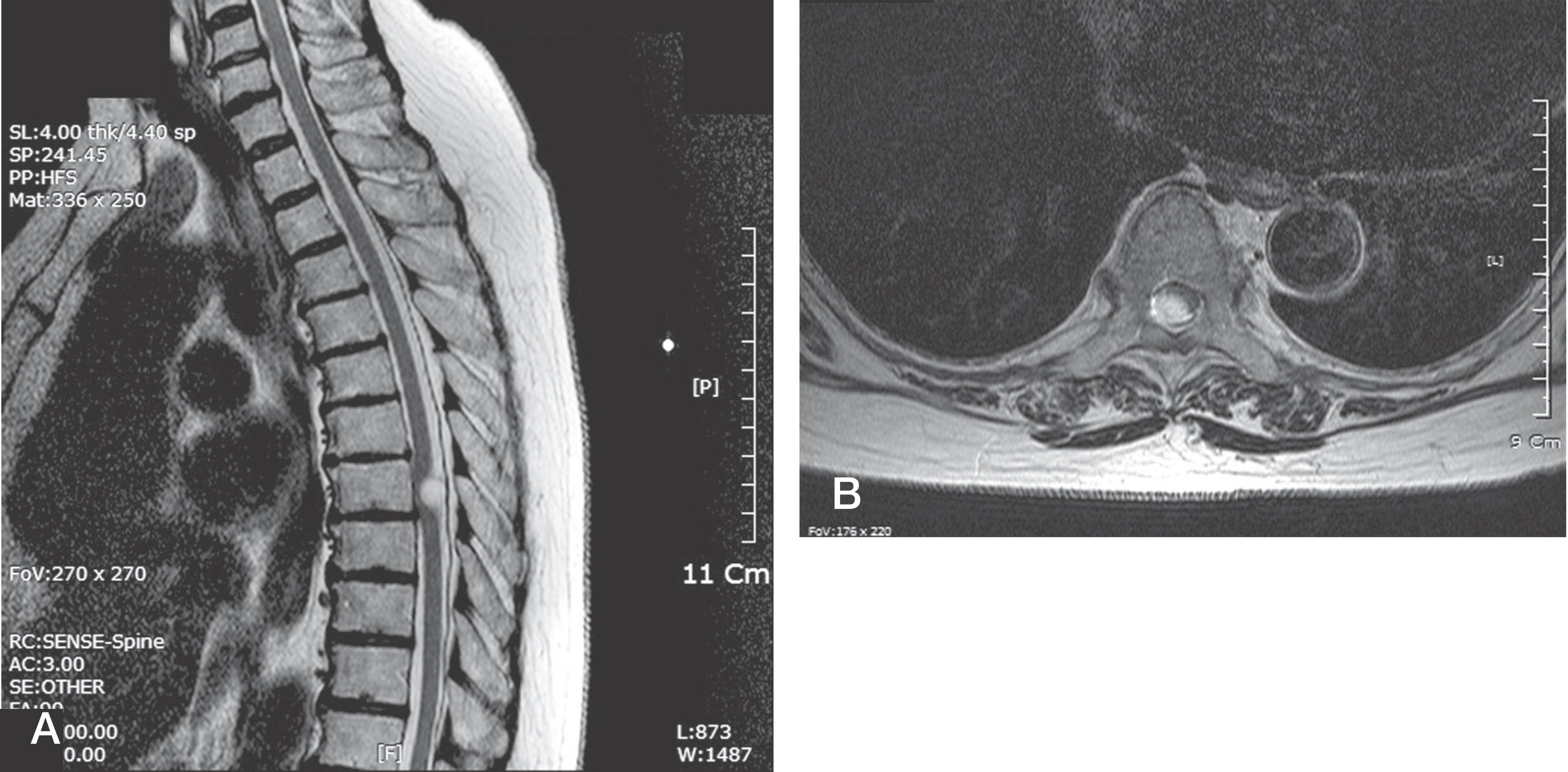
Fig. 1. MRI finding of lumbar spine (A) Cystic mass with hyper-intense signal area in T2 weighted image at the T7. (B) Tumor mass occupies the almost entire canal in axial views.
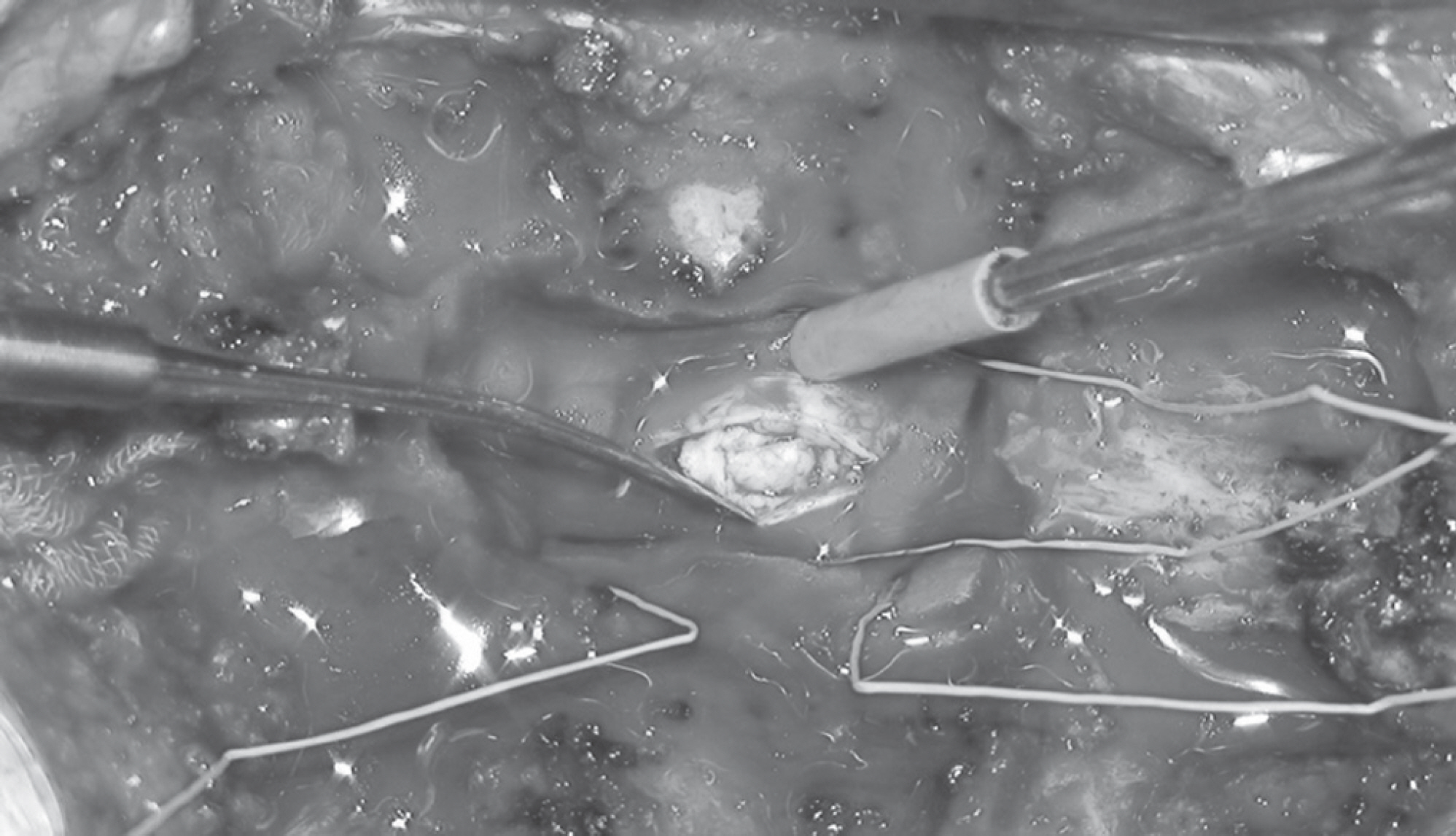
Fig. 2. Operative Findings : Huge irregularly shaped nodular tumor mass wall is seen, when the dura was open.
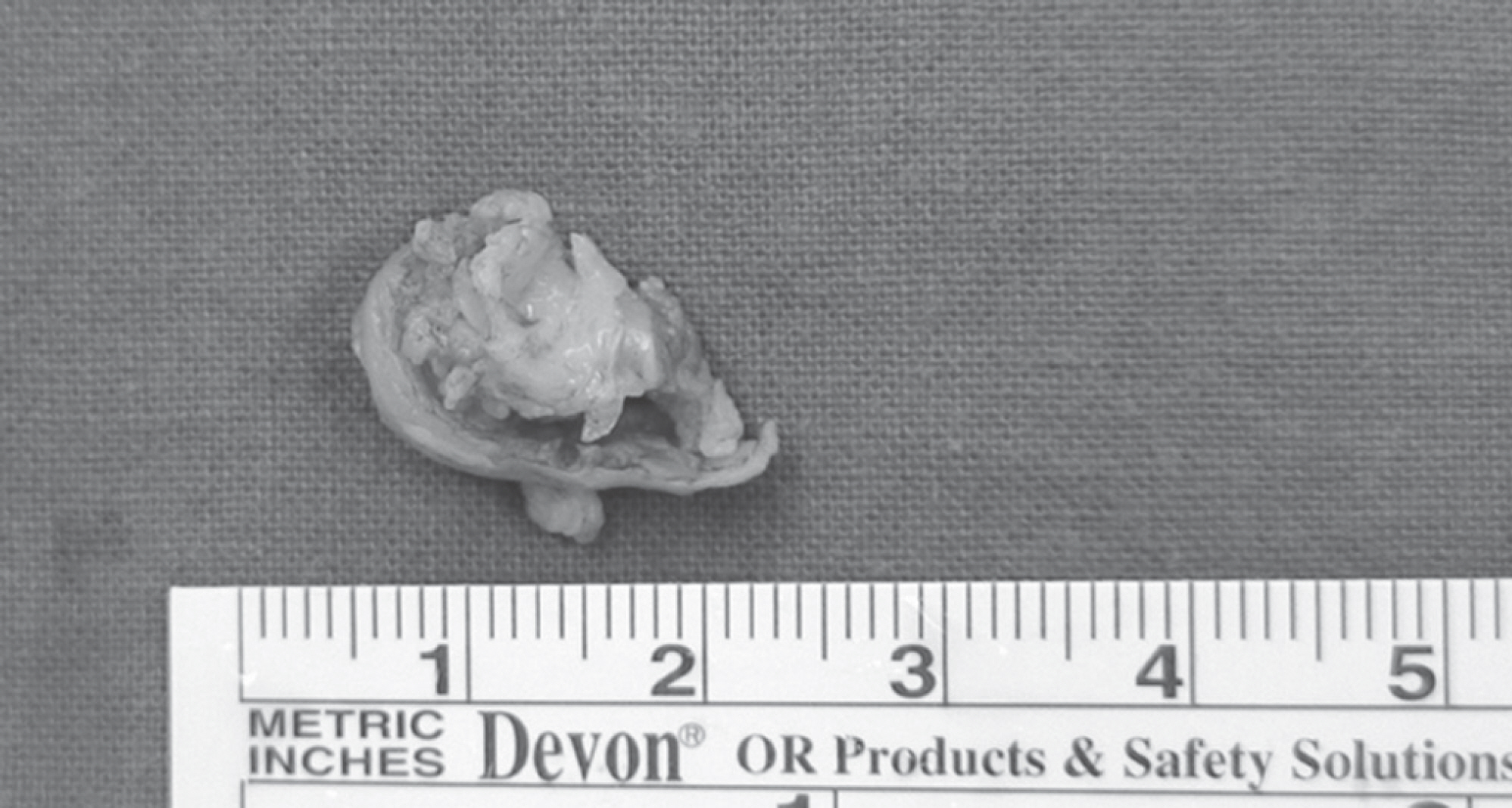
Fig. 3. Operative Findings : 1x1.5 size mass is excised.
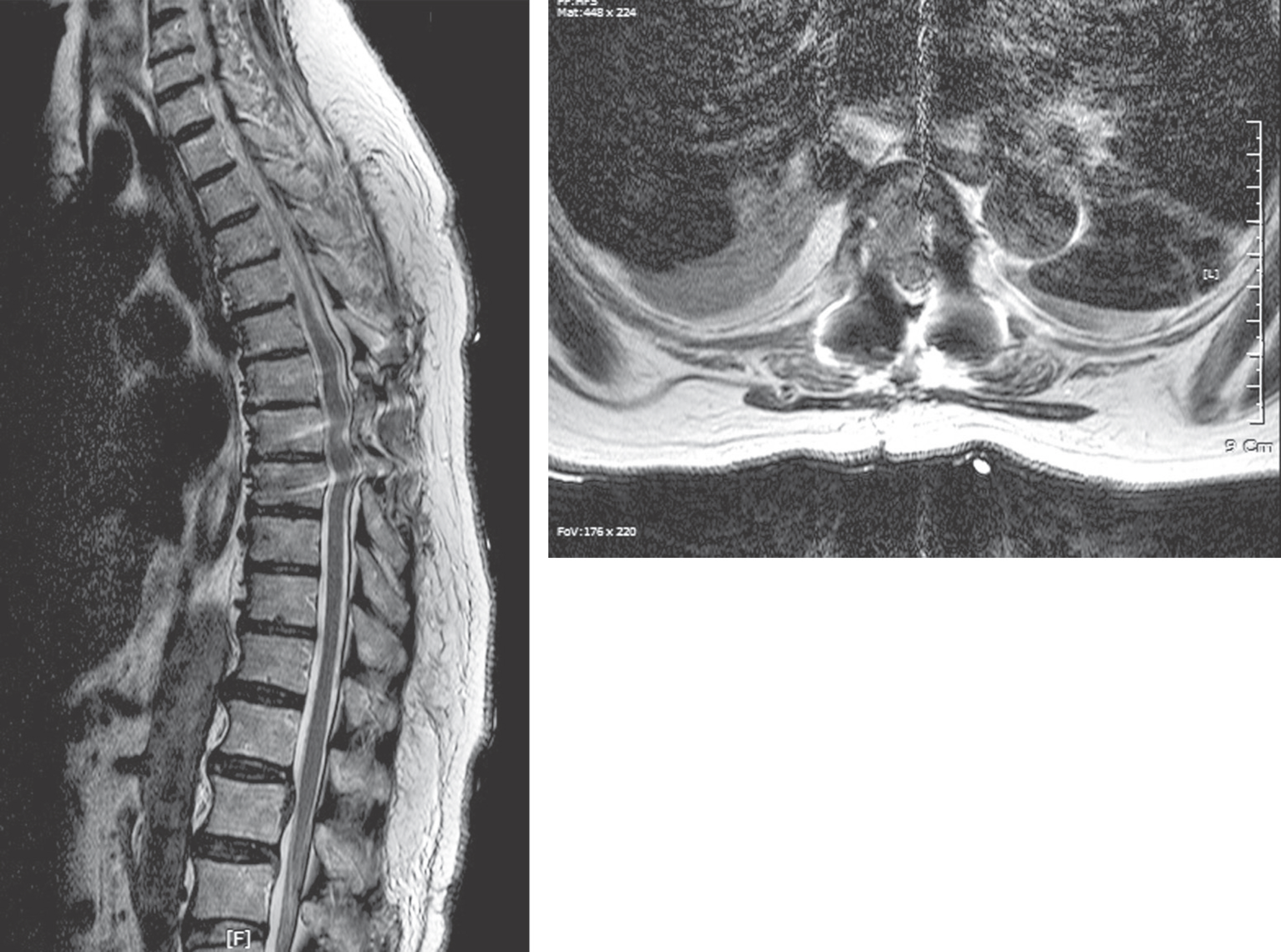
Fig. 4. Sagittal and axial magnetic resonance images 1 weeks after surgery reveal normalization of the previously compressed spinal cord.
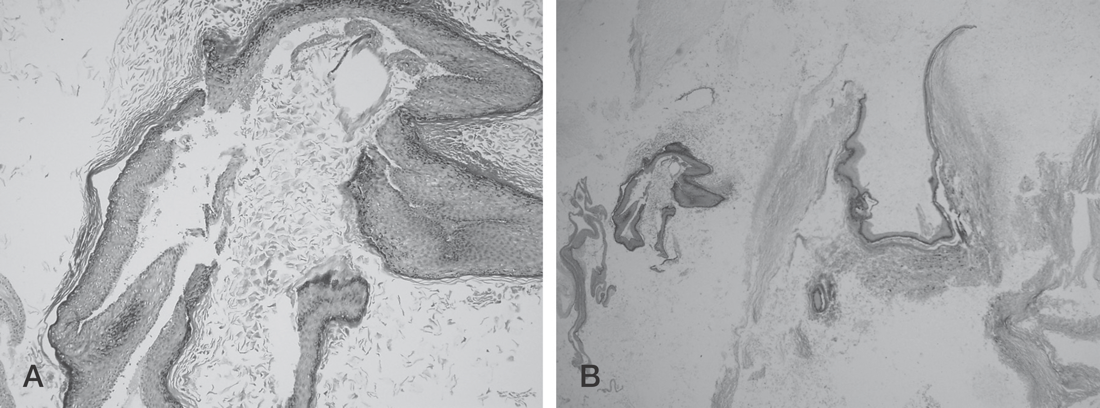
Fig. 5. (A) The microscopic finding shows keratin laid down by the well-developed squamous epithelium. The stroma reveals abun-dant skin appendages. But no sweat gland and hair follicles are seen. (B) The well-developed squamous epithelia are shown. The underlying stroma demonstrates loose mesenchymal tissue(x100).
Reference
-
1. Alvisi C, Cerisoli M, Giulioni M, Guerra L. Longterm results of surgically treated congenital intradural spinal arachnoid cysts. J Neurosurg. 1987; 67:333–5.
Article2. Manno NJ, Uihlein A, Kernohan JW. Intraspinal epider-moids. J Neurosurg. 1962; 19:754–65.
Article3. Alves AM, Norrell H. Intramedullary epidermoid tumors of the spinal cord. Report of a case and review of the literature. Int Surg. 1970; 54:239–43.4. Roux A, Mercier C, Larbrisseau A, Dube LJ, Dupuis C, Del Carpio R. Intramedullary epidermoid cysts of the spinal cord. Case report. J Neurosurg. 1992; 76:528–33.5. Hatfield MK, Udesky RH, Strimling AM, Kim BH, Sibergleit R. MR imaging of a spinal epidermoid tumor. AJNR Am J Neuroradiol. 1989; 10:S95–6.6. Lai SW, Chan WP, Chen CY, Chien JC, Chu JS, Chiu WT. MRI of epidermoid cyst of the conus medullaris. Spinal Cord. 2005; 43:320–3.
Article7. Horowitz BL, Chari MV, James R, Bryan RN. MR of intracranial epidermoid tumors: correlation of in vivo imaging with in vitro 13C spectroscopy. AJNR Am J Neuroradiol. 1990; 11:299–302.8. Dunn RC Jr., Archer CA, Rapport RL 2nd, Looi LM. Unusual CT-dense posterior fossa epidermoid cyst: case report. J Neurosurg. 1981; 55:654–6.9. Debray MP, Ricolfi F, Brugieres P, Khalil A, Adle-Biassette H, Gaston A. Epidermoid cyst of the conus medullaris: atypical MRI and angiographic features. Neuroradiology. 1996; 38:526–8.
Article10. Cervoni L, Celli P, Cantore G, Fortuna A. Intradural tumors of the cauda equina: a single institution review of clinical characteristics. Clin Neurol Neurosurg. 1995; 97:8–12.
Article
- Full Text Links
-
- Actions
-
Cited
- CITED
-
- Close
- Share
-
- Similar articles
-
- Extensive Intradural Epidermoid Cysts with Cauda Equina Syndrome in the Lumbosacral Spine: Case Report
- Lumbar Epidermoid Inclusion Cyst after Single Spinal Puncture: A case report
- A Rare Case of Thoracic Intradural Epidermoid Cyst after Spinal Cord Stimulator Insertion: A Case Report
- Intradural Epidermoid Cyst: A Case Report
- Multiple Intramedullary and Intradural Epidermoid Cysts in the Conus Medullaris and the Lumbar Spine: Case Report
