J Korean Med Sci.
2009 Feb;24(1):179-183. 10.3346/jkms.2009.24.1.179.
Bowel Obstruction Caused by an Intramural Duodenal Hematoma: A Case Report of Endoscopic Incision and Drainage
- Affiliations
-
- 1Department of Internal Medicine, Bundang CHA Hospital, College of Medicine, Pochon CHA University, Seongnam, Korea. mdkwon71@freechal.com
- KMID: 1794430
- DOI: http://doi.org/10.3346/jkms.2009.24.1.179
Abstract
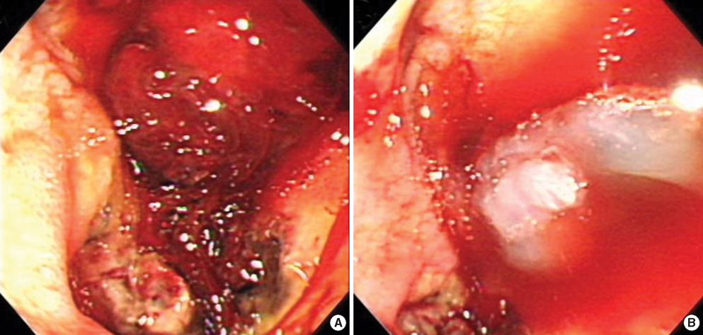
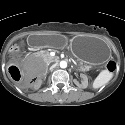
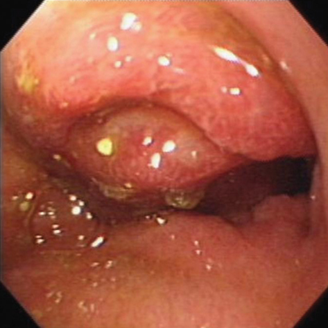
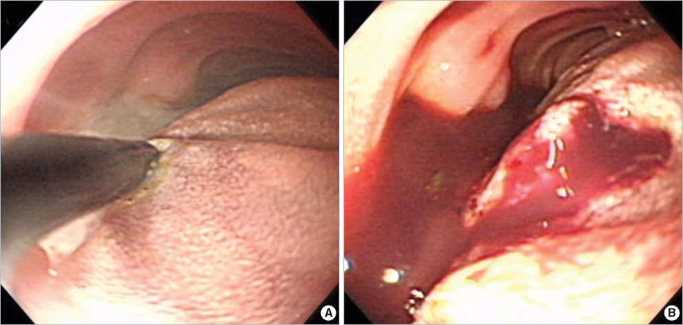
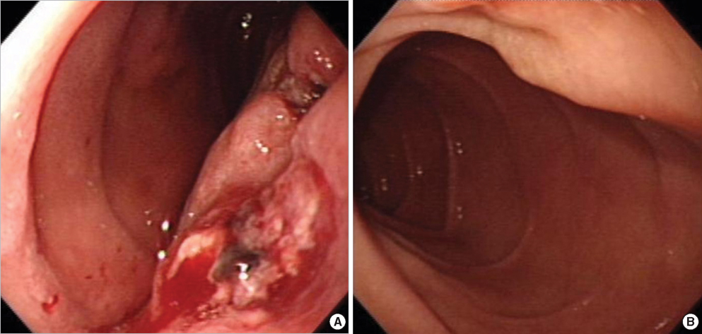
- Complications associated with an intramural hematoma of the bowel, is a relatively unusual condition. Most intramural hematomas resolve spontaneously with conservative treatment and the patient prognosis is good. However, if the symptoms are not resolved or the condition persists, surgical intervention may be necessary. Here we describe internal incision and drainage by endoscopy for the treatment of an intramural hematoma of the duodenum. A 63-yr-old woman was admitted to the hospital with hematemesis. The esophagogastroduodenoscopy (EGD) showed active ulcer bleeding at the distal portion of duodenal bulb. A total of 10 mL of 0.2% epinephrine and 2 mL of fibrin glue were injected locally. The patient developed diffuse abdominal pain and projectile vomiting three days after the endoscopic treatment. An abdominal computed tomography revealed a very large hematoma at the lateral duodenal wall, approximately 10X5 cm in diameter. Follow-up EGD was performed showing complete luminal obstruction at the second portion of the duodenum caused by an intramural hematoma. The patient's condition was not improved with conservative treatment. Therefore, 21 days after admission, endoscopic treatment of the hematoma was attempted. Puncture and incision were performed with an electrical needle knife. Two days after the procedure, the patient was tolerating a soft diet without complaints of abdominal pain or vomiting. The hematoma resolved completely on the follow-up studies.
Keyword
MeSH Terms
Figure
Cited by 2 articles
-
Huge Intramural Duodenal Hematoma Complicated with Obstructive Jaundice following Endoscopic Hemostasis
Hak Su Kim, Hee Kyoung Kim, Won Hee Kim, Sung Pyo Hong, Joo Young Cho
Korean J Gastroenterol. 2019;73(1):39-44. doi: 10.4166/kjg.2019.73.1.39.Acute Duodenal Ischemia and Periampullary Intramural Hematoma after an Uneventful Endoscopic Retrograde Cholangiopancreatography in a Patient with Primary Myelofibrosis
Chang Ho Jung, Jong Jin Hyun, Dae Hoe Gu, Eul Sun Moon, Jae Seon Kim, Hong Sik Lee, Chang Duck Kim
Clin Endosc. 2014;47(3):270-274. doi: 10.5946/ce.2014.47.3.270.
Reference
-
1. Hughes CE 3rd, Conn J Jr, Sherman JO. Intramural hematoma of the gastrointestinal tract. Am J Surg. 1977. 133:276–279.
Article2. Ghishan FK, Werner M, Vieira P, Kuttesch J, DeHaro R. Intramural duodenal hematoma: an unusual complication of endoscopic small bowel biopsy. Am J Gastroenterol. 1987. 82:368–370.3. Zinelis SA, Hershenson LM, Ennis MF, Boller M, Ismail-Beigi F. Intramural duodenal hematoma following upper gastrointestinal endoscopic biopsy. Dig Dis Sci. 1989. 34:289–291.
Article4. Rohrer B, Schreiner J, Lehnert P, Waldner H, Heldwein W. Gastrointestinal intramural hematoma, a complication of endoscopic injection methods for bleeding peptic ulcers: a case series. Endoscopy. 1994. 26:617–621.
Article5. Lipson SA, Perr HA, Koerper MA, Ostroff JW, Snyder JD, Goldstein RB. Intramural duodenal hematoma after endoscopic biopsy in leukemic patients. Gastrointest Endosc. 1996. 44:620–623.
Article6. Guzman C, Bousvaros A, Buonomo C, Nurko S. Intraduodenal hematoma complicating intestinal biopsy: case reports and review of the literature. Am J Gastroenterol. 1998. 93:2547–2550.
Article7. Yen HH, Soon MS, Chen YY. Esophageal intramural hematoma: an unusual complication of endoscopic biopsy. Gastrointest Endosc. 2005. 62:161–163.
Article8. Sugai K, Kajiwara E, Mochizuki Y, Noma E, Nakashima J, Uchimura K, Sadoshima S. Intramural duodenal hematoma after endoscopic therapy for a bleeding duodenal ulcer in a patient with liver cirrhosis. Intern Med. 2005. 44:954–957.
Article9. Beal JM, Haid S, Sherrick J, Scarff J, Rambach W, Method H. Small bowel obstruction secondary to hematoma of the mesentery. IMJ Ill Med J. 1966. 130:422–426.10. Jewett TC Jr, Caldarola V, Karp MP, Allen JE, Cooney DR. Intramural hematoma of the duodenum. Arch Surg. 1988. 123:54–58.
Article11. Polat C, Dervisoglu A, Guven H, Kaya E, Malazgirt Z, Danaci M, Ozkan K. Anticoagulant-induced intramural intestinal hematoma. Am J Emerg Med. 2003. 21:208–211.
Article12. Aizawa K, Tokuyama H, Yonezawa T, Doi M, Matsuzono Y, Matumoto M, Uragami K, Nishioka S, Yataka I. A case report of traumatic intramural hematoma of the duodenum effectively treated with ultrasonically guided aspiration drainage and endoscopic balloon catheter dilation. Gastroenterol Jpn. 1991. 26:218–223.13. Maemura T, Yamaguchi Y, Yukioka T, Matsuda H, Shimazaki S. Laparoscopic drainage of an intramural duodenal hematoma. J Gastroenterol. 1999. 34:119–122.
Article14. Murata N, Kuroda T, Fujino S, Murata M, Takagi S, Seki M. Submucosal dissection of the esophagus: a case report. Endoscopy. 1991. 23:95–97.
Article15. Bak YT, Kwon OS, Yeon JE, Kim JS, Byun KS, Kim JH, Kim JG, Lee CH, Choi YH, Kang DH. Endoscopic treatment in a case with extensive spontaneous intramural dissection of the oesophagus. Eur J Gastroenterol Hepatol. 1998. 10:969–972.
Article16. Cho CM, Ha SS, Tak WY, Kweon YO, Kim SK, Choi YH, Chung JM. Endoscopic incision of a septum in a case of spontaneous intramural dissection of the esophagus. J Clin Gastroenterol. 2002. 35:387–390.
Article17. K C S, Kouzu T, Matsutani S, Hishikawa E, Saisho H. Early endoscopic treatment of intramural hematoma of the esophagus. Gastrointest Endosc. 2003. 58:297–301.18. Adachi T, Togashi H, Watanabe H, Okumoto K, Hattori E, Takeda T, Terui Y, Aoki M, Ito J, Sugahara K, Saito K, Saito T, Kawata S. Endoscopic incision for esophageal intramural hematoma after injection sclerotherapy: case report. Gastrointest Endosc. 2003. 58:466–468.
- Full Text Links
- Actions
-
Cited
- CITED
-
- Close
- Share
- Similar articles
-
- Successful Endoscopic Decompression for Intramural Duodenal Hematoma with Gastric Outlet Obstruction Complicating Acute Pancreatitis
- Intramural Hematoma of the Duodenum following Endoscopic Biopsy in a Child with Henoch-Schonlein Purpura
- Huge Intramural Duodenal Hematoma Complicated with Obstructive Jaundice following Endoscopic Hemostasis
- A Rare Cause of Duodenal Obstruction: Spontaneous Intramural Duodenal Hematoma Caused by a Hemangioma
- A Case of Intramural Duodenal Hematoma after the Use of the Endoscopic Epinephrine Injection Method for Duodenal Ulcer Bleeding in a Chronic Renal Failure Patient undergoing Maintenance Hemodialysis

