Huge Intramural Duodenal Hematoma Complicated with Obstructive Jaundice following Endoscopic Hemostasis
- Affiliations
-
- 1Department of Gastroenterology and Hepatology, Incheon Sarang Hospital, Incheon, Korea.
- 2Department of Gastroenterology and Hepatology, Cheonggu Sungsim Hospital, Seoul, Korea.
- 3Digestive Disease Center, CHA Bundang Medical Center, CHA University, Seongnam, Korea. ari98@cha.ac.kr
- KMID: 2432435
- DOI: http://doi.org/10.4166/kjg.2019.73.1.39
Abstract
- Intramural hematoma of the duodenum is a relatively unusual complication associated with the endoscopic treatment of bleeding peptic ulcers. Intramural hematomas are typically resolved spontaneously with conservative treatment alone. We report a case of an intramural duodenal hematoma following endoscopic hemostasis with epinephrine injection therapy, which was associated with transient obstructive jaundice in a patient undergoing hemodialysis. The patient developed biliary sepsis due to obstruction of the common bile duct secondary to the huge hematoma. He was treated with fluoroscopy-guided drainage catheter insertion, which spontaneously resolved the biliary sepsis through conservative treatment in 6 weeks. Fluoroscopy-guided drainage may impact the treatment of intramural hematomas that involve life-threatening complications.
Keyword
MeSH Terms
Figure
-
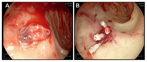
Fig. 1 Endoscopic findings. (A) The esophago-gastro-duodenoscopy on admission revealed a deep ulcer and an exposed vessel on the base was noted at the duodenal bulb. (B) An epinephrine injection (2 mL, 1 mL) was administered to control the blood oozing from the vessel. Complete hemostasis was acquired with four hemoclippings and 2 mL of fibrin glue injection.
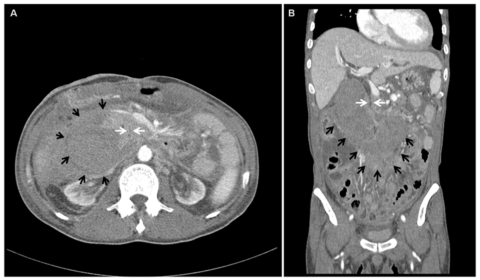
Fig. 2 On the 2nd hospital day, the contrast-enhanced abdominal computed tomography scan showed a huge intramural hematoma in the 2nd to 4th portions of the duodenum with compression of the common bile duct (black arrows, intramural hematoma; white arrows, common bile duct). (A) Axial image. (B) Coronal image.

Fig. 3 Fluoroscopic finding. An 8-Fr pigtail catheter was inserted through the right transhepatic access to the duodenal submucosal hematoma.
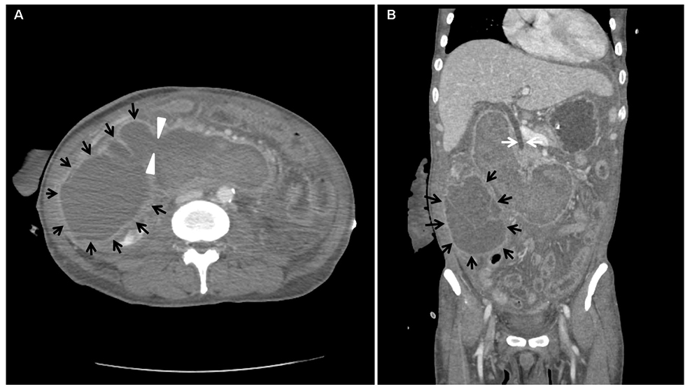
Fig. 4 Six days after catheter insertion, the follow-up abdominal computed tomography showed partial regression of the intramural hematoma and mild improvement of the common bile duct obstruction. A marked increase in loculated fluid collection was shown in the right abdomen with fistula formation leading to the retroperitoneum from the duodenal submucosal hematoma (black arrows, hematoma fluid loculation; arrowheads, fistula; white arrows, common bile duct). (A) Axial image. (B) Coronal image.
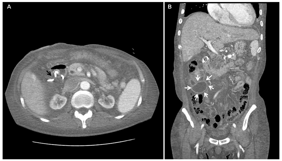
Fig. 5 After 4 weeks of catheter drainage, the follow-up abdominal computed tomography showed near resorption of the hematoma in the submucosal area of the duodenum and decreased loculated fluid collection in the right abdomen (black arrow, resolved intramural hematoma; white arrows, decreased loculated fluid). (A) Axial image. (B) Coronal image.

Fig. 6 Esophago-gastro-duodenoscopy at 8 weeks revealed the healed duodenal ulcer without any evidence of hematoma.
Reference
-
1. Rohrer B, Schreiner J, Lehnert P, Waldner H, Heldwein W. Gastrointestinal intramural hematoma, a complication of endoscopic injection methods for bleeding peptic ulcers: a case series. Endoscopy. 1994; 26:617–621.
Article2. Dhawan V, Mohamed A, Fedorak RN. Gastric intramural hematoma: a case report and literature review. Can J Gastroenterol. 2009; 23:19–22.
Article3. Laine L, McQuaid KR. Endoscopic therapy for bleeding ulcers: an evidence-based approach based on meta-analyses of randomized controlled trials. Clin Gastroenterol Hepatol. 2009; 7:33–47. quiz 1–2.
Article4. Chung S, Park CW, Chung HW, Shin SJ, Chang YS. Intramural duodenal hematoma and hemoperitoneum after endoscopic treatment in a patient with chronic renal failure on hemodialysis: a case report. Cases J. 2009; 2:9083.
Article5. Sugai K, Kajiwara E, Mochizuki Y, et al. Intramural duodenal hematoma after endoscopic therapy for a bleeding duodenal ulcer in a patient with liver cirrhosis. Intern Med. 2005; 44:954–957.
Article6. Kwon CI, Choi KH, Ko EH, et al. A case of duodenal intramural hematoma treated by percutaneous external drainage. Korean J Gastroenterol. 2007; 49:45–49.7. Zinelis SA, Hershenson LM, Ennis MF, Boller M, Ismail-Beigi F. Intramural duodenal hematoma following upper gastrointestinal endoscopic biopsy. Dig Dis Sci. 1989; 34:289–291.
Article8. Kwon CI, Ko KH, Kim HY, et al. Bowel obstruction caused by an intramural duodenal hematoma: a case report of endoscopic incision and drainage. J Korean Med Sci. 2009; 24:179–183.
Article9. Plojoux O, Hauser H, Wettstein P. Computed tomography of intramural hematoma of the small intestine: a report of 3 cases. Radiology. 1982; 144:559–561.
Article10. Aizawa K, Tokuyama H, Yonezawa T, et al. A case of traumatic intramural hematoma of the duodenum effectively treated with ultrasonically guided aspiration drainage and endoscopic balloon catheter dilation. Gastroenterol Jpn. 1991; 26:218–223.
- Full Text Links
-
- Actions
-
Cited
- CITED
-
- Close
- Share
-
- Similar articles
-
- A Case of Intramural Duodenal Hematoma Complicated with Obstructive Jaundice and Pancreatitis following Endoscopic Hemostasis
- Acute Cholecystitis and Obstructive Jaundice by Nontraumatic Duodenal Intramural Hematoma at Ampulla of Vater
- Intramural Duodenal Hematoma following Endoscopic Epinephrine and Thrombin Injection for Bleeding Duodenal Ulcer in a Geriatric Patient with a History of Anticoagulant Drug Use
- Intramural Duodenal Hematoma Complicated with Pancreatitis after Endoscopic Hemostasis in a Chronic Renal Failure Patient with Maintenance Hemodialysis
- A Case of Intramural Duodenal Hematoma Accompanied by Acute Pancreatitis Following Endoscopic Hemostasis for Duodenal Ulcer Bleeding
