Stent Evaluation with Optical Coherence Tomography
- Affiliations
-
- 1Division of Cardiology, Severance Cardiovascular Hospital, Yonsei University College of Medicine, Seoul, Korea. mkhong61@yuhs.ac
- 2Severance Biomedical Science Institute, Yonsei University College of Medicine, Seoul, Korea.
- KMID: 1793152
- DOI: http://doi.org/10.3349/ymj.2013.54.5.1075
Abstract
- Optical coherence tomography (OCT) has been recently applied to investigate coronary artery disease in interventional cardiology. Compared to intravascular ultrasound, OCT is able to visualize various vascular structures more clearly with higher resolution. Several validation studies have shown that OCT is more accurate in evaluating neointimal tissue after coronary stent implantation than intravascular ultrasound. Novel findings on OCT evaluation include the detection of strut coverage and the characterization of neointimal tissue in an in-vivo setting. In a previous study, neointimal healing of stent strut was pathologically the most important factor associated with stent thrombosis, a fatal complication, in patients treated with drug-eluting stent (DES). Recently, OCT-defined coverage of a stent strut was proposed to be related with clinical safety in DES-treated patients. Neoatherosclerosis is an atheromatous change of neointimal tissue within the stented segment. Clinical studies using OCT revealed neoatherosclerosis contributed to late-phase luminal narrowing after stent implantation. Like de novo native coronary lesions, the clinical presentation of OCT-derived neoatherosclerosis varied from stable angina to acute coronary syndrome including late stent thrombosis. Thus, early identification of neoatherosclerosis with OCT may predict clinical deterioration in patients treated with coronary stent. Additionally, intravascular OCT evaluation provides additive information about the performance of coronary stent. In the near future, new advances in OCT technology will help reduce complications with stent therapy and accelerating in the study of interventional cardiology.
MeSH Terms
Figure
-
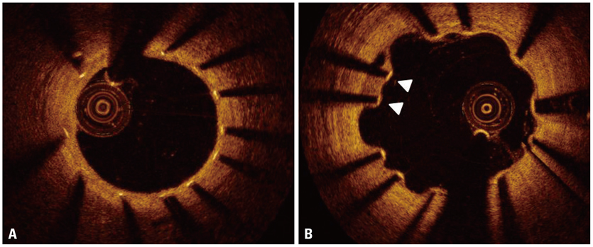
Fig. 1 Representative images of well-apposed vs. malapposed struts. Optical coherence tomography shows well-apposed struts with complete coverage 9 months after drug-eluting stent implantation (A), whereas some struts (arrowheads) show incomplete stent apposition and uncovered portions to the lumen (B).

Fig. 2 Strut coverage of drug-eluting stent (DES) over time. A dot represents each of the studies in Table 1, except for Bayesian hierarchical models. A curved line represents estimated change of uncovered strut after DES implantation. Usage of sirolimus-eluting stent and incomplete stent apposition increases the risk of delayed coverage after DES implantation.
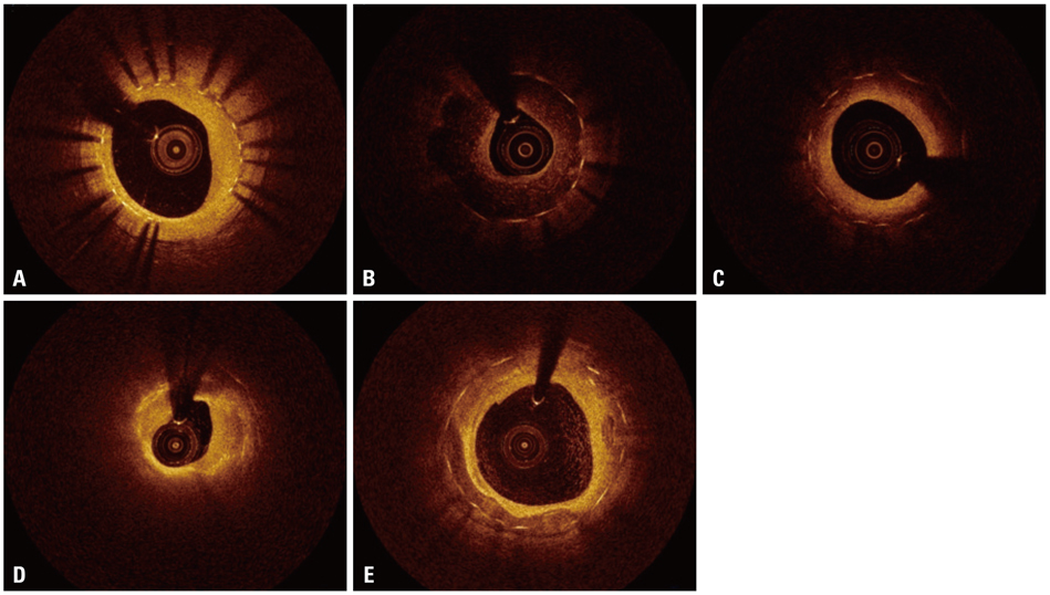
Fig. 3 Various patterns of neointimal tissue. (A) Homogeneous pattern, (B) heterogeneous pattern, (C) layered pattern, (D) lipid-laden neointima, (E) neointima with calcification.
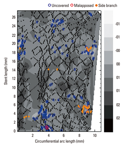
Fig. 4 Contour plot of strut coverage after drug-eluting stent implantation, using optical coherence tomography. The contour plot shows detailed information about the position and coverage of stent strut, in which the circumferential arc is plotted along the X axis and the stent length is plotted along the Y axis. Blue circles represent uncovered struts, red circles represent malapposed struts, and orange circles represent struts crossing over a side-branch vessel.53
Cited by 3 articles
-
Prospective and Systematic Analysis of Unexpected Requests for Non-Cardiac Surgery or Other Invasive Procedures during the First Year after Drug-Eluting Stent Implantation
Byeong-Keuk Kim, Jung-Han Yoon, Dong-Ho Shin, Jung-Sun Kim, Young-Guk Ko, Donghoon Choi, Seung-Hwan Lee, Gary S. Mintz, Yangsoo Jang, Myeong-Ki Hong,
Yonsei Med J. 2014;55(2):345-352. doi: 10.3349/ymj.2014.55.2.345.Optical Coherence Tomographic Observation of Morphological Features of Neointimal Tissue after Drug-Eluting Stent Implantation
Seung-Yul Lee, Dong-Ho Shin, Jung-Sun Kim, Byeong-Keuk Kim, Young-Guk Ko, Donghoon Choi, Yangsoo Jang, Myeong-Ki Hong
Yonsei Med J. 2014;55(4):944-952. doi: 10.3349/ymj.2014.55.4.944.Impact of Statin Treatment on Strut Coverage after Drug-Eluting Stent Implantation
Yongsung Suh, Byeong-Keuk Kim, Dong-Ho Shin, Jung-Sun Kim, Young-Guk Ko, Donghoon Choi, Yangsoo Jang, Myeong-Ki Hong
Yonsei Med J. 2015;56(1):45-52. doi: 10.3349/ymj.2015.56.1.45.
Reference
-
1. Serruys PW, de Jaegere P, Kiemeneij F, Macaya C, Rutsch W, Heyndrickx G, et al. Benestent Study Group. A comparison of balloon-expandable-stent implantation with balloon angioplasty in patients with coronary artery disease. N Engl J Med. 1994; 331:489–495.
Article2. Fischman DL, Leon MB, Baim DS, Schatz RA, Savage MP, Penn I, et al. Stent Restenosis Study Investigators. A randomized comparison of coronary-stent placement and balloon angioplasty in the treatment of coronary artery disease. N Engl J Med. 1994; 331:496–501.
Article3. Forrester JS, Fishbein M, Helfant R, Fagin J. A paradigm for restenosis based on cell biology: clues for the development of new preventive therapies. J Am Coll Cardiol. 1991; 17:758–769.
Article4. Virmani R, Farb A. Pathology of in-stent restenosis. Curr Opin Lipidol. 1999; 10:499–506.
Article5. Moses JW, Leon MB, Popma JJ, Fitzgerald PJ, Holmes DR, O'Shaughnessy C, et al. Sirolimus-eluting stents versus standard stents in patients with stenosis in a native coronary artery. N Engl J Med. 2003; 349:1315–1323.
Article6. Stone GW, Ellis SG, Cox DA, Hermiller J, O'Shaughnessy C, Mann JT, et al. A polymer-based, paclitaxel-eluting stent in patients with coronary artery disease. N Engl J Med. 2004; 350:221–231.
Article7. Daemen J, Wenaweser P, Tsuchida K, Abrecht L, Vaina S, Morger C, et al. Early and late coronary stent thrombosis of sirolimus-eluting and paclitaxel-eluting stents in routine clinical practice: data from a large two-institutional cohort study. Lancet. 2007; 369:667–678.
Article8. Wenaweser P, Daemen J, Zwahlen M, van Domburg R, Jüni P, Vaina S, et al. Incidence and correlates of drug-eluting stent thrombosis in routine clinical practice. 4-year results from a large 2-institutional cohort study. J Am Coll Cardiol. 2008; 52:1134–1140.
Article9. Suzuki Y, Ikeno F, Koizumi T, Tio F, Yeung AC, Yock PG, et al. In vivo comparison between optical coherence tomography and intravascular ultrasound for detecting small degrees of in-stent neointima after stent implantation. JACC Cardiovasc Interv. 2008; 1:168–173.
Article10. Kwon SW, Kim BK, Kim TH, Kim JS, Ko YG, Choi D, et al. Qualitative assessment of neointimal tissue after drug-eluting stent implantation: comparison between follow-up optical coherence tomography and intravascular ultrasound. Am Heart J. 2011; 161:367–372.
Article11. Takarada S, Imanishi T, Liu Y, Ikejima H, Tsujioka H, Kuroi A, et al. Advantage of next-generation frequency-domain optical coherence tomography compared with conventional time-domain system in the assessment of coronary lesion. Catheter Cardiovasc Interv. 2010; 75:202–206.
Article12. Bezerra HG, Attizzani GF, Sirbu V, Musumeci G, Lortkipanidze N, Fujino Y, et al. Optical coherence tomography versus intravascular ultrasound to evaluate coronary artery disease and percutaneous coronary intervention. JACC Cardiovasc Interv. 2013; 6:228–236.
Article13. Nakano M, Vorpahl M, Otsuka F, Taniwaki M, Yazdani SK, Finn AV, et al. Ex vivo assessment of vascular response to coronary stents by optical frequency domain imaging. JACC Cardiovasc Imaging. 2012; 5:71–82.
Article14. Hong MK, Mintz GS, Lee CW, Park DW, Park KM, Lee BK, et al. Late stent malapposition after drug-eluting stent implantation: an intravascular ultrasound analysis with long-term follow-up. Circulation. 2006; 113:414–419.
Article15. Tearney GJ, Regar E, Akasaka T, Adriaenssens T, Barlis P, Bezerra HG, et al. Consensus standards for acquisition, measurement, and reporting of intravascular optical coherence tomography studies: a report from the International Working Group for Intravascular Optical Coherence Tomography Standardization and Validation. J Am Coll Cardiol. 2012; 59:1058–1072.16. Kim WH, Lee BK, Lee S, Shim JM, Kim JS, Kim BK, et al. Serial changes of minimal stent malapposition not detected by intravascular ultrasound: follow-up optical coherence tomography study. Clin Res Cardiol. 2010; 99:639–644.
Article17. Kim JS, Kim JS, Shin DH, Kim BK, Ko YG, Choi D, et al. Optical coherence tomographic comparison of neointimal coverage between sirolimus- and resolute zotarolimus-eluting stents at 9 months after stent implantation. Int J Cardiovasc Imaging. 2012; 28:1281–1287.
Article18. Choi HH, Kim JS, Yoon DH, Hong KS, Kim TH, Kim BK, et al. Favorable neointimal coverage in everolimus-eluting stent at 9 months after stent implantation: comparison with sirolimus-eluting stent using optical coherence tomography. Int J Cardiovasc Imaging. 2012; 28:491–497.
Article19. Gutiérrez-Chico JL, Jüni P, García-García HM, Regar E, Nüesch E, Borgia F, et al. Long-term tissue coverage of a biodegradable polylactide polymer-coated biolimus-eluting stent: comparative sequential assessment with optical coherence tomography until complete resorption of the polymer. Am Heart J. 2011; 162:922–931.
Article20. Katoh H, Shite J, Shinke T, Matsumoto D, Tanino Y, Ogasawara D, et al. Delayed neointimalization on sirolimus-eluting stents: 6-month and 12-month follow up by optical coherence tomography. Circ J. 2009; 73:1033–1037.
Article21. Gutiérrez-Chico JL, Wykrzykowska J, Nüesch E, van Geuns RJ, Koch KT, Koolen JJ, et al. Vascular tissue reaction to acute malapposition in human coronary arteries: sequential assessment with optical coherence tomography. Circ Cardiovasc Interv. 2012; 5:20–29. S1–S8.
Article22. Kawamori H, Shite J, Shinke T, Otake H, Matsumoto D, Nakagawa M, et al. Natural consequence of post-intervention stent malapposition, thrombus, tissue prolapse, and dissection assessed by optical coherence tomography at mid-term follow-up. Eur Heart J Cardiovasc Imaging. 2013; [Epub ahead of print].
Article23. Gutiérrez-Chico JL, Regar E, Nüesch E, Okamura T, Wykrzykowska J, di Mario C, et al. Delayed coverage in malapposed and side-branch struts with respect to well-apposed struts in drug-eluting stents: in vivo assessment with optical coherence tomography. Circulation. 2011; 124:612–623.
Article24. Kim BK, Hong MK, Shin DH, Kim JS, Ko YG, Choi D, et al. Relationship between stent malapposition and incomplete neointimal coverage after drug-eluting stent implantation. J Interv Cardiol. 2012; 25:270–277.
Article25. Kim BK, Shin DH, Kim JS, Ko YG, Choi D, Jang Y, et al. Optical coherence tomography-based evaluation of malapposed strut coverage after drug-eluting stent implantation. Int J Cardiovasc Imaging. 2012; 28:1887–1894.
Article26. Kim JS, Hong MK, Shin DH, Kim BK, Ko YG, Choi D, et al. Quantitative and qualitative changes in DES-related neointimal tissue based on serial OCT. JACC Cardiovasc Imaging. 2012; 5:1147–1155.
Article27. Takano M, Yamamoto M, Mizuno M, Murakami D, Inami T, Kimata N, et al. Late vascular responses from 2 to 4 years after implantation of sirolimus-eluting stents: serial observations by intracoronary optical coherence tomography. Circ Cardiovasc Interv. 2010; 3:476–483.
Article28. Finn AV, Joner M, Nakazawa G, Kolodgie F, Newell J, John MC, et al. Pathological correlates of late drug-eluting stent thrombosis: strut coverage as a marker of endothelialization. Circulation. 2007; 115:2435–2441.
Article29. Matsumoto D, Shite J, Shinke T, Otake H, Tanino Y, Ogasawara D, et al. Neointimal coverage of sirolimus-eluting stents at 6-month follow-up: evaluated by optical coherence tomography. Eur Heart J. 2007; 28:961–967.
Article30. Kim JS, Kim JS, Kim TH, Fan C, Lee JM, Kim W, et al. Comparison of neointimal coverage of sirolimus-eluting stents and paclitaxel-eluting stents using optical coherence tomography at 9 months after implantation. Circ J. 2010; 74:320–326.
Article31. Kim JS, Jang IK, Kim JS, Kim TH, Takano M, Kume T, et al. Optical coherence tomography evaluation of zotarolimus-eluting stents at 9-month follow-up: comparison with sirolimus-eluting stents. Heart. 2009; 95:1907–1912.
Article32. Kim BK, Hong MK, Shin DH, Nam CM, Kim JS, Ko YG, et al. A new strategy for discontinuation of dual antiplatelet therapy: the RESET Trial (REal Safety and Efficacy of 3-month dual antiplatelet Therapy following Endeavor zotarolimus-eluting stent implantation). J Am Coll Cardiol. 2012; 60:1340–1348.33. Sarno G, Lagerqvist B, Fröbert O, Nilsson J, Olivecrona G, Omerovic E, et al. Lower risk of stent thrombosis and restenosis with unrestricted use of 'new-generation' drug-eluting stents: a report from the nationwide Swedish Coronary Angiography and Angioplasty Registry (SCAAR). Eur Heart J. 2012; 33:606–613.
Article34. Barlis P, Regar E, Serruys PW, Dimopoulos K, van der Giessen WJ, van Geuns RJ, et al. An optical coherence tomography study of a biodegradable vs. durable polymer-coated limus-eluting stent: a LEADERS trial sub-study. Eur Heart J. 2010; 31:165–176.
Article35. Stefanini GG, Kalesan B, Serruys PW, Heg D, Buszman P, Linke A, et al. Long-term clinical outcomes of biodegradable polymer biolimus-eluting stents versus durable polymer sirolimus-eluting stents in patients with coronary artery disease (LEADERS): 4 year follow-up of a randomised non-inferiority trial. Lancet. 2011; 378:1940–1948.
Article36. Guagliumi G, Sirbu V, Musumeci G, Gerber R, Biondi-Zoccai G, Ikejima H, et al. Examination of the in vivo mechanisms of late drug-eluting stent thrombosis: findings from optical coherence tomography and intravascular ultrasound imaging. JACC Cardiovasc Interv. 2012; 5:12–20.
Article37. Won H, Shin DH, Kim BK, Mintz GS, Kim JS, Ko YG, et al. Optical coherence tomography derived cut-off value of uncovered stent struts to predict adverse clinical outcomes after drug-eluting stent implantation. Int J Cardiovasc Imaging. 2013; [Epub ahead of print].
Article38. Kim BK, Kim JS, Park J, Ko YG, Choi D, Jang Y, et al. Comparison of optical coherence tomographic assessment between first- and second-generation drug-eluting stents. Yonsei Med J. 2012; 53:524–529.
Article39. Gonzalo N, Serruys PW, Okamura T, van Beusekom HM, Garcia-Garcia HM, van Soest G, et al. Optical coherence tomography patterns of stent restenosis. Am Heart J. 2009; 158:284–293.
Article40. Lee SJ, Kim BK, Kim JS, Ko YG, Choi D, Jang Y, et al. Evaluation of neointimal morphology of lesions with or without in-stent restenosis: an optical coherence tomography study. Clin Cardiol. 2011; 34:633–639.
Article41. Nakazawa G, Otsuka F, Nakano M, Vorpahl M, Yazdani SK, Ladich E, et al. The pathology of neoatherosclerosis in human coronary implants bare-metal and drug-eluting stents. J Am Coll Cardiol. 2011; 57:1314–1322.42. Prati F, Regar E, Mintz GS, Arbustini E, Di Mario C, Jang IK, et al. Expert review document on methodology, terminology, and clinical applications of optical coherence tomography: physical principles, methodology of image acquisition, and clinical application for assessment of coronary arteries and atherosclerosis. Eur Heart J. 2010; 31:401–415.
Article43. Yonetsu T, Kim JS, Kato K, Kim SJ, Xing L, Yeh RW, et al. Comparison of incidence and time course of neoatherosclerosis between bare metal stents and drug-eluting stents using optical coherence tomography. Am J Cardiol. 2012; 110:933–939.
Article44. Yonetsu T, Kato K, Kim SJ, Xing L, Jia H, McNulty I, et al. Predictors for neoatherosclerosis: a retrospective observational study from the optical coherence tomography registry. Circ Cardiovasc Imaging. 2012; 5:660–666.45. Habara M, Terashima M, Nasu K, Kaneda H, Inoue K, Ito T, et al. Difference of tissue characteristics between early and very late restenosis lesions after bare-metal stent implantation: an optical coherence tomography study. Circ Cardiovasc Interv. 2011; 4:232–238.
Article46. Habara M, Terashima M, Nasu K, Kaneda H, Yokota D, Ito T, et al. Morphological differences of tissue characteristics between early, late, and very late restenosis lesions after first generation drug-eluting stent implantation: an optical coherence tomography study. Eur Heart J Cardiovasc Imaging. 2013; 14:276–284.
Article47. Kimura T, Yokoi H, Nakagawa Y, Tamura T, Kaburagi S, Sawada Y, et al. Three-year follow-up after implantation of metallic coronary-artery stents. N Engl J Med. 1996; 334:561–566.
Article48. Collet CA, Costa JR, Abizaid A, Chamié D, Staico R, Costa R, et al. Assessing the temporal course of neointimal hyperplasia formation after different generations of drug-eluting stents. JACC Cardiovasc Interv. 2011; 4:1067–1074.
Article49. Kang SJ, Mintz GS, Akasaka T, Park DW, Lee JY, Kim WJ, et al. Optical coherence tomographic analysis of in-stent neoatherosclerosis after drug-eluting stent implantation. Circulation. 2011; 123:2954–2963.
Article50. Ko YG, Kim DM, Cho JM, Choi SY, Yoon JH, Kim JS, et al. Optical coherence tomography findings of very late stent thrombosis after drug-eluting stent implantation. Int J Cardiovasc Imaging. 2012; 28:715–723.
Article51. Nagai H, Ishibashi-Ueda H, Fujii K. Histology of highly echolucent regions in optical coherence tomography images from two patients with sirolimus-eluting stent restenosis. Catheter Cardiovasc Interv. 2010; 75:961–963.
Article52. Okamura T, Onuma Y, García-García HM, Regar E, Wykrzykowska JJ, Koolen J, et al. 3-Dimensional optical coherence tomography assessment of jailed side branches by bioresorbable vascular scaffolds: a proposal for classification. JACC Cardiovasc Interv. 2010; 3:836–844.
Article53. Ha J, Kim BK, Kim JS, Shin DH, Ko YG, Choi D, et al. Assessing neointimal coverage after DES implantation by 3D OCT. JACC Cardiovasc Imaging. 2012; 5:852–853.
Article54. Giattina SD, Courtney BK, Herz PR, Harman M, Shortkroff S, Stamper DL, et al. Assessment of coronary plaque collagen with polarization sensitive optical coherence tomography (PS-OCT). Int J Cardiol. 2006; 107:400–409.
Article55. Tahara N, Imaizumi T, Virmani R, Narula J. Clinical feasibility of molecular imaging of plaque inflammation in atherosclerosis. J Nucl Med. 2009; 50:331–334.
Article56. Kim JS, Jang IK, Fan C, Kim TH, Kim JS, Park SM, et al. Evaluation in 3 months duration of neointimal coverage after zotarolimus-eluting stent implantation by optical coherence tomography: the ENDEAVOR OCT trial. JACC Cardiovasc Interv. 2009; 2:1240–1247.
Article57. Kim S, Kim JS, Shin DH, Kim BK, Ko YG, Choi D, et al. Comparison of early strut coverage between zotarolimus- and everolimus-eluting stents using optical coherence tomography. Am J Cardiol. 2013; 111:1–5.
Article58. Gutiérrez-Chico JL, van Geuns RJ, Regar E, van der Giessen WJ, Kelbæk H, Saunamäki K, et al. Tissue coverage of a hydrophilic polymer-coated zotarolimus-eluting stent vs. a fluoropolymer-coated everolimus-eluting stent at 13-month follow-up: an optical coherence tomography substudy from the RESOLUTE All Comers trial. Eur Heart J. 2011; 32:2454–2463.
Article59. Inoue T, Shite J, Yoon J, Shinke T, Otake H, Sawada T, et al. Optical coherence evaluation of everolimus-eluting stents 8 months after implantation. Heart. 2011; 97:1379–1384.
Article60. Takano M, Murakami D, Yamamoto M, Kurihara O, Murai K, Inami T, et al. Six-month follow-up evaluation for everolimus-eluting stents by intracoronary optical coherence tomography: comparison with paclitaxel-eluting stents. Int J Cardiol. 2013; 166:181–186.
Article61. Kim TH, Kim JS, Kim BK, Ko YG, Choi D, Jang Y, et al. Long-term (≥2 years) follow-up optical coherence tomographic study after sirolimus- and paclitaxel-eluting stent implantation: comparison to 9-month follow-up results. Int J Cardiovasc Imaging. 2011; 27:875–881.
Article
- Full Text Links
-
- Actions
-
Cited
- CITED
-
- Close
- Share
-
- Similar articles
-
- Seven Fractures in Three Second Generation Drug Eluting Stents Implanted in the Right Coronary Artery Assessed by Using Optical Coherence Tomography
- A Newly Developed Stent Thrombus Related to Optical Coherence Tomography
- Identification of Vulnerable Plaque in a Stented Coronary Segment 17 Years after Implantation Using Optical Coherence Tomography
- Availability of Optical Coherence Tomography in Diagnosis and Classification of Choroidal Neovascularization
- A Newly Formed and Ruptured Atheromatous Plaque within Neointima after Drug-Eluting Stent Implantation: 2-Year Follow-Up Intravascular Ultrasound and Optical Coherence Tomography Studies
