Korean J Gastroenterol.
2012 Jun;59(6):433-436. 10.4166/kjg.2012.59.6.433.
Diagnosis of an Accessory Spleen Mimicking a Gastric Submucosal Tumor Using Endoscopic Ultrasonography-guided Fine-needle Aspiration
- Affiliations
-
- 1Department of Gastroenterology, Asan Medical Center, University of Ulsan College of Medicine, Seoul, Korea. hyjung@amc.seoul.kr
- 2Department of Pathology, Asan Medical Center, University of Ulsan College of Medicine, Seoul, Korea.
- KMID: 1792849
- DOI: http://doi.org/10.4166/kjg.2012.59.6.433
Abstract
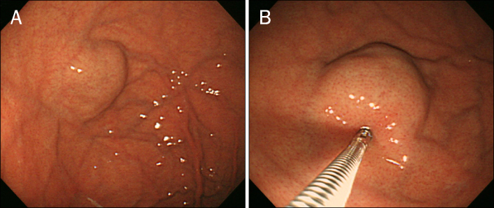
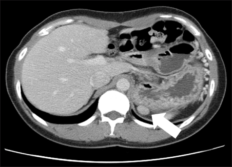
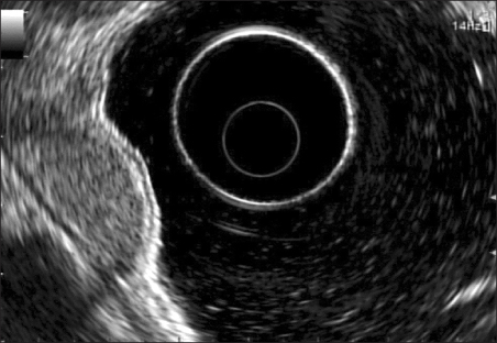
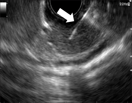
- Accessory spleen can be mistaken as a gastric subepithelial mass, and may not be differentiated in CT or endoscopic ultrasonography (EUS). A gastric subepithelial mass was detected on routine endoscopy in a 39-year old woman with history of splenectomy. In subsequent CT and EUS, the subepithelial mass was located on the fourth layer of the stomach. To make a definite diagnosis, EUS-guided fine needle aspiration (FNA) was performed, and a splenic tissue was demonstrated in histologic examination. EUS-guided FNA can be beneficial in the diagnosis of accessory spleen which mimics a gastric subepithelial mass.
Keyword
MeSH Terms
Figure
Reference
-
1. Beahrs JR, Stephens DH. Enlarged accessory spleens: CT appearance in postsplenectomy patients. AJR Am J Roentgenol. 1980. 135:483–486.2. Halpert B, Gyorkey F. Lesions observed in accessory spleens of 311 patients. Am J Clin Pathol. 1959. 32:165–168.3. Curtis GM, Movitz D. The surgical significance of the accessory spleen. Ann Surg. 1946. 123:276–298.4. Verheyden CN, Beart RW Jr, Clifton MD, Phyliky RL. Accessory splenectomy in management of recurrent idiopathic thrombocytopenic purpura. Mayo Clin Proc. 1978. 53:442–446.5. Coote JM, Eyers PS, Walker A, Wells IP. Intra-abdominal bleeding caused by spontaneous rupture of an accessory spleen: the CT findings. Clin Radiol. 1999. 54:689–691.6. Valls C, Monés L, Gumà A, López-Calonge E. Torsion of a wandering accessory spleen: CT findings. Abdom Imaging. 1998. 23:194–195.7. Seo T, Ito T, Watanabe Y, Umeda T. Torsion of an accessory spleen presenting as an acute abdomen with an inflammatory mass. US, CT, and MRI findings. Pediatr Radiol. 1994. 24:532–534.8. Brewster DC. Splenosis. Report of two cases and review of the literature. Am J Surg. 1973. 126:14–19.9. Chin S, Isomoto H, Mizuta Y, Wen CY, Shikuwa S, Kohno S. Enlarged accessory spleen presenting stomach submucosal tumor. World J Gastroenterol. 2007. 13:1752–1754.10. Gayer G, Zissin R, Apter S, Atar E, Portnoy O, Itzchak Y. CT findings in congenital anomalies of the spleen. Br J Radiol. 2001. 74:767–772.11. Balthazar EJ. CT of the gastrointestinal tract: principles and interpretation. AJR Am J Roentgenol. 1991. 156:23–32.12. Kim JY, Lee JM, Kim KW, et al. Ectopic pancreas: CT findings with emphasis on differentiation from small gastrointestinal stromal tumor and leiomyoma. Radiology. 2009. 252:92–100.13. Motoo Y, Okai T, Ohta H, et al. Endoscopic ultrasonography in the diagnosis of extraluminal compressions mimicking gastric submucosal tumors. Endoscopy. 1994. 26:239–242.14. Yoshida S, Yamashita K, Yokozawa M, et al. Diagnostic findings of ultrasound-guided fine-needle aspiration cytology for gastrointestinal stromal tumors: proposal of a combined cytology with newly defined features and histology diagnosis. Pathol Int. 2009. 59:712–719.15. Chatzipantelis P, Salla C, Karoumpalis I, et al. Endoscopic ultrasound-guided fine needle aspiration biopsy in the diagnosis of gastrointestinal stromal tumors of the stomach. A study of 17 cases. J Gastrointestin Liver Dis. 2008. 17:15–20.16. Schreiner AM, Mansoor A, Faigel DO, Morgan TK. Intrapancreatic accessory spleen: mimic of pancreatic endocrine tumor diagnosed by endoscopic ultrasound-guided fine-needle aspiration biopsy. Diagn Cytopathol. 2008. 36:262–265.17. Fritscher-Ravens A, Mylonaki M, Pantes A, Topalidis T, Thonke F, Swain P. Endoscopic ultrasound-guided biopsy for the diagnosis of focal lesions of the spleen. Am J Gastroenterol. 2003. 98:1022–1027.
- Full Text Links
- Actions
-
Cited
- CITED
-
- Close
- Share
- Similar articles
-
- Endoscopic Ultrasound-Guided Fine Needle Aspiration in Submucosal Lesion
- Endoscopic Fine Needle Aspiration Cytology in the Diagnosis of Upper Gastrointestinal Malignancies
- Tuberculous Lymphadenitis Mimicking Gastric Subepithelial Tumor Diagnosed Using Endoscopic Ultrasound-guided Fine-needle Aspiration
- Fine-Needle Biopsy: Should This Be the First Choice in Endoscopic Ultrasound-Guided Tissue Acquisition?
- Hepatic Tuberculosis Granuloma Mimicking Hilar Cholangiocarcinoma Confirmed by Endoscopic Ultrasound-Guided Fine-Needle Aspiration


