Characteristics of Advanced Gastric Cancer Undetected on Gastroscopy
- Sung IK
- Kim YC
- Yun JW
- Seo HI

- Park DI
- Cho YK
- Kim HJ
- Park JH
- Sohn CI
- Jeon WK
- Kim BI
- Oh SJ
- Son BH
- Yoo CH
- Sohn JH
- Lee HY
- Won KH
- Affiliations
-
- 1Department of Internal Medicine, Konkuk University Medical Center, Seoul, Korea.
- 2Department of Internal Medicine, Kangbuk Samsung Hospital, Sungkyunkwan University, Seoul, Korea. chongil.sohn@samsung.com
- 3Department of Oncology, Kangbuk Samsung Hospital, Sungkyunkwan University, Seoul, Korea.
- 4Department of Surgery, Kangbuk Samsung Hospital, Sungkyunkwan University, Seoul, Korea.
- 5Department of Pathology, Kangbuk Samsung Hospital, Sungkyunkwan University, Seoul, Korea.
- 6Department of Internal Medicine, CHA University School of Medicine, Seoul, Korea.
- 7Health Screening Center, Kangbuk Samsung Hospital, Sungkyunkwan University School of Medicine, Suwon, Korea.
- KMID: 1792785
- DOI: http://doi.org/10.4166/kjg.2011.57.5.288
Abstract
- BACKGROUND/AIMS
Stomach cancer can be easily diagnosed via endoscopy, but also possible to be missed. The aim of this study was to investigate the clinical and endoscopic characteristics of advanced gastric cancers that were not diagnosed based on endoscopic examination.
METHODS
We evaluated patients who had newly diagnosed advanced gastric cancer that was undetected via endoscopy within the last six months.
RESULTS
Sixteen patients were included in this study. The locations of the cancers were the cardia in six cases, the greater curvature side of the body in eight cases and the antrum in two cases. The histological findings were tubular type adenocarcinoma in 11 cases, with ten cases of moderately to poorly differentiated adenocarcinoma and five cases of signet ring cell type adenocarcinoma.
CONCLUSIONS
Even advanced gastric cancer lesions may not be detected during endoscopy. If a patient continues to complain of upper gastrointestinal symptoms, even though endoscopy does not find abnormal findings, repeated endoscopy and/or additional diagnostic studies should be considered.
Keyword
MeSH Terms
Figure
-
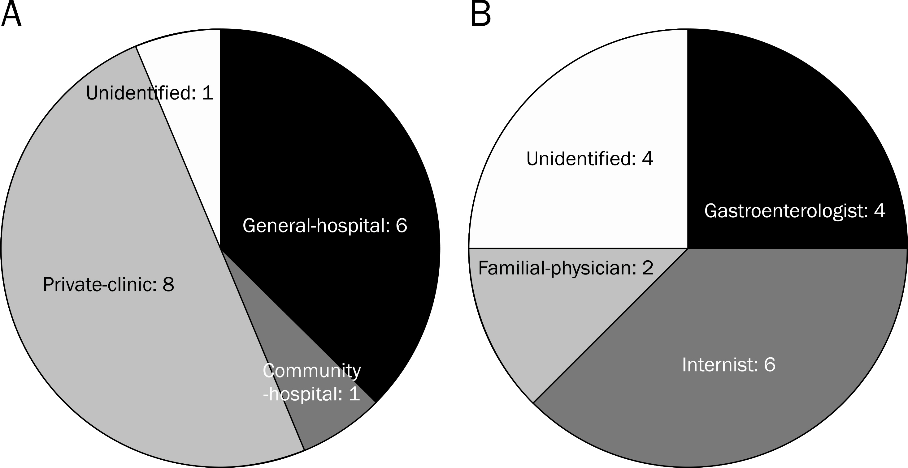
Fig. 1. Hospitals and endoscopists of prior endoscopic examinations which could not detect gastric cancer. (A) Most of the hospitals were general hospitals or private clinics. (B) Most of the prior endoscopists were gastroenterologists or internists.
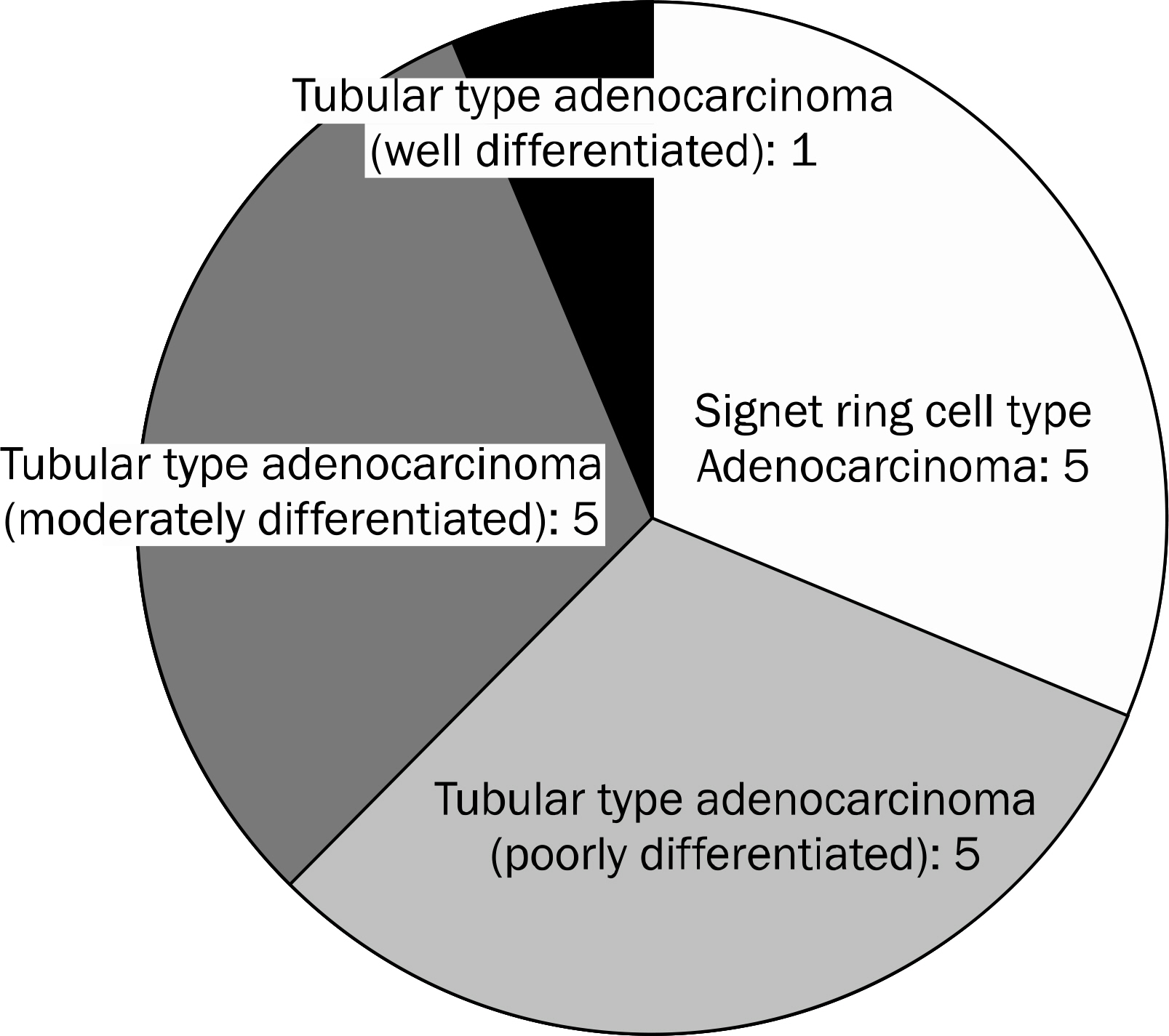
Fig. 2. Histological characteristics of misdiagnosed advanced gastric cancer.

Fig. 3. Endoscopic and histological classifications of misdiagnosed advanced gastric cancer according to the location in the stomach. (A) Most cancers were located around the cardia and greater curvature side of the body. Endoscopically, the cancers around the cardia are Borrmann type III, and the cancers on the greater curvature side of the body were Borrmann type IV. (B) Microscopically, the cancers around the cardia were moderately differentiated, and the cancers on the greater curvature side of body were signet ring cell and poorly differentiated.
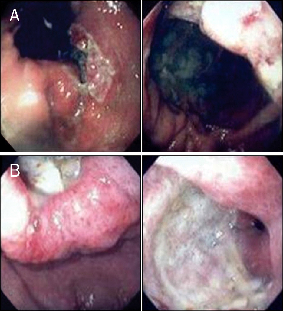
Fig. 4. Endoscopic finding of misdiagnosed advanced gastric cancer around the cardia (A) and antrum (B). (A) The endoscopic findings on later examination showed a large ulceroinfiltrative lesion around the cardia. (B) A very large ulcerative lesion was observed on the second endoscopic examination. The prior endoscopist misdiagnosed the encircling antral ulcerative cancer lesion as a duodenal ulcer within the duodenal bulb.

Fig. 5. Endoscopic finding of misdiagnosed advanced gastric cancer on the greater curvature side of the body. (A) On the first endoscopic examination, there was no remarkable lesion on the greater curvature side of body except for an ulcer scar on the duodenal bulb. (B) However, three months later, endoscopic examination showed a very large irregularly shaped ulcer with thickened mucosal folds and poor expansibility along the greater curvature side of stomach from the antrum up to the mid-body.
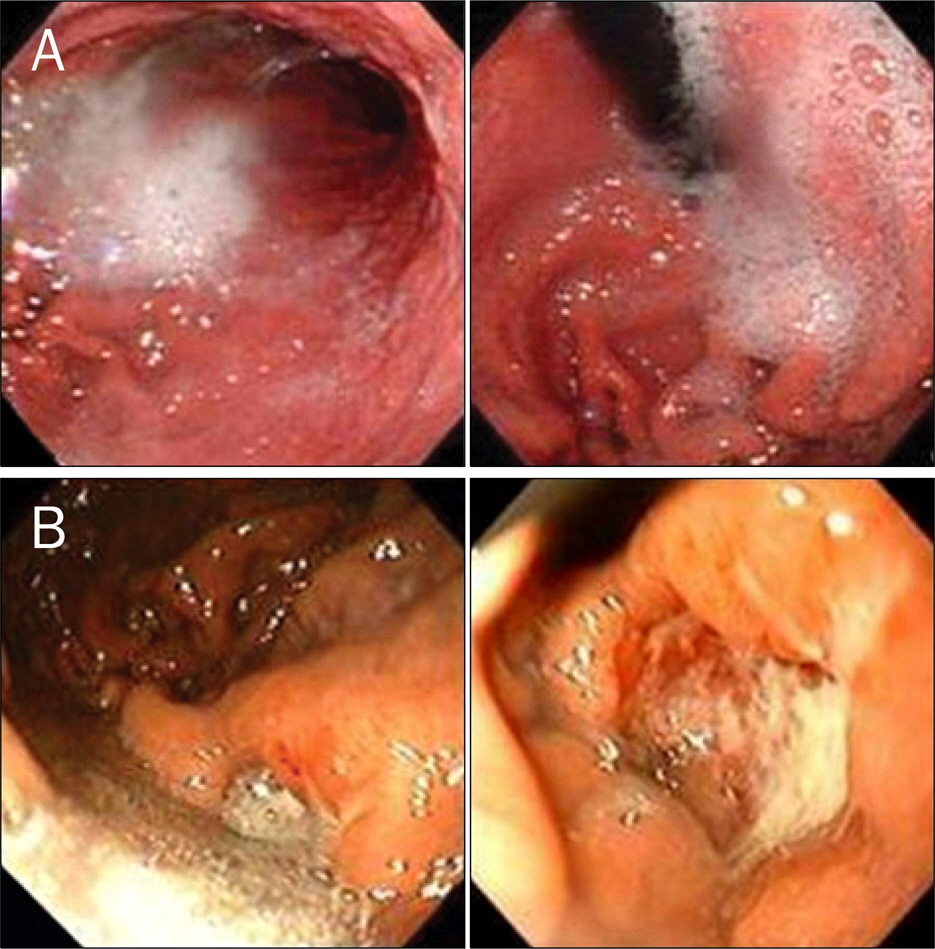
Fig. 6. Endoscopic finding of misdiagnosed advanced gastric cancer of the upper body. (A) On the previous endoscopic photograph, there appeared to be no abnormal lesion; there were gastric secretions and air bubbles on the greater curvature side of the upper body. (B) Follow-up endoscopic examination revealed a large round ulcerative cancer lesion on the greater curvature side of the upper body. This lesion was missed because of inadequate removal of gastric secretions during the first endoscopic examination.
Cited by 2 articles
-
Characteristics of Missed Simultaneous Gastric Lesions Based on Double-Check Analysis of the Endoscopic Image
Eun Jeong Gong, Jeong Hoon Lee, Kyoungwon Jung, Charles J. Cho, Hee Kyong Na, Ji Yong Ahn, Kee Wook Jung, Do Hoon Kim, Kee Don Choi, Ho June Song, Gin Hyug Lee, Hwoon-Yong Jung, Jin-Ho Kim
Clin Endosc. 2017;50(3):261-269. doi: 10.5946/ce.2016.056.Characteristics of Missed Synchronous Gastric Epithelial Neoplasms
Bong Eun Lee
Clin Endosc. 2017;50(3):211-212. doi: 10.5946/ce.2017.058.
Reference
-
References
1. Ngoan LT, Yoshimura T. Pattern and time trends of stomach cancer in Asia from 1950–99. Asian Pac J Cancer Prev. 2002; 3:47–54.2. Kokkola A, Sipponen P. Gastric carcinoma in young adults. Hepatogastroenterology. 2001; 48:1552–1555.3. Eguchi T, Takahashi Y, Yamagata M, Kasahara M, Fujii M. Gastric cancer in young patients. J Am Coll Surg. 1999; 188:22–26.
Article4. Axon AT, Bell GD, Jones RH, Quine MA, McCloy RF. Guidelines on appropriate indications for upper gastrointestinal endoscopy. Working Party of the Joint Committee of the Royal College of Physicians of London, Royal College of Surgeons of England, Royal College of Anaesthetists, Association of Surgeons, the British Society of Gastroenterology, and the Thoracic Society of Great Britain. BMJ. 1995; 310:853–856.5. Hallissey MT, Allum WH, Jewkes AJ, Ellis DJ, Fielding JW. Early detection of gastric cancer. BMJ. 1990; 301:513–515.
Article6. Graham DY, Schwartz JT, Cain GD, Gyorkey F. Prospective evaluation of biopsy number in the diagnosis of esophageal and gastric carcinoma. Gastroenterology. 1982; 82:228–231.
Article7. Siersema PD, Dees J, Tilanus HW, Kok TC, Hordijk ML, van Blankenstein M. Early detection and treatment of oesophageal and gastric cancer. The Rotterdam Oesophageal Tumour Study Group. Neth J Med. 1995; 47:76–86.8. Hosokawa O, Tsuda S, Kidani E, et al. Diagnosis of gastric cancer up to three years after negative upper gastrointestinal endoscopy. Endoscopy. 1998; 30:669–674.
Article9. An-Foraker SH, Vise D. Cytodiagnosis of gastric carcinoma, linitis plastica type (diffuse, infiltrating, poorly differentiated adenocarcinoma). Acta Cytol. 1981; 25:361–366.10. Levine MS, Kong V, Rubesin SE, Laufer I, Herlinger H. Scirrhous carcinoma of the stomach: radiologic and endoscopic diagnosis. Radiology. 1990; 175:151–154.
Article11. Ming SC, Goldman H. Pathology of the gastrointestinal tract. Philadelphia: WB Saunders;1992.12. el-Zimaity HM, Itani K, Graham DY. Early diagnosis of signet ring cell carcinoma of the stomach: role of the Genta stain. J Clin Pathol. 1997; 50:867–868.
Article13. Minami M, Kawauchi N, Itai Y, Niki T, Sasaki Y. Gastric tumors: radiologic-pathologic correlation and accuracy of T staging with dynamic CT. Radiology. 1992; 185:173–178.
Article14. Kuntz C, Herfarth C. Imaging diagnosis for staging of gastric cancer. Semin Surg Oncol. 1999; 17:96–102.
Article15. Murakami R, Tsukuma H, Ubukata T, et al. Estimation of validity of mass screening program for gastric cancer in Osaka, Japan. Cancer. 1990; 65:1255–1260.
Article16. Flatau E, Resnitzky P, Grishkan A, Chaimowich O, Levy E. Linitis plastica infiltrating the entire gut. Am J Gastroenterol. 1982; 77:559–561.17. Kanter MA, Isaacson NH, Knoll SM, Nochomovitz LE. The diagnostic challenge of metastatic linitis plastica. Two cases and a consideration of the problem. Am Surg. 1986; 52:510–513.18. Nakajima T, Konishi H, Tatsumi Y, et al. Gastric cancer presenting with extremely rapid growth: unprecedented morphologic change in a short time and endoscopic estimation of its doubling time. Endoscopy. 2000; 32:994–997.
Article
- Full Text Links
-
- Actions
-
Cited
- CITED
-
- Close
- Share
-
- Similar articles
-
- Virtual Gastroscopy
- The Endoscopic Findings of Gastric Lymphoma
- Gastric Cancer After Bariatric Surgeries
- A study on the life style, locus of control and health belief of gastric cancer patients
- The Clinicopathologic Features and Recurrence of Resection-Line Involvement of Gastric Cancer after Gastrectomy
