Treatment of an 8-mm Myxoma Using Acellular Corneal Tissue
- Affiliations
-
- 1Department of Ophthalmology, Chung-Ang University College of Medicine, Seoul, Korea. jck50ey@kornet.net
- KMID: 1792098
- DOI: http://doi.org/10.3341/kjo.2014.28.1.86
Abstract
- A myxoma is a benign tumor found in the heart and in various soft tissues; however, a corneal myxoma is rare. A mucinous mass of unknown etiology was observed on the left cornea of a 32-year-old male patient. We performed deep anterior lamellar keratoplasty using acellular corneal tissue and concurrent amniotic membrane transplantation. Hematoxylin and eosin staining revealed vacuolation of the parenchyma and myxoid change in the corneal tissue that occurred in the anterior half of the corneal parenchyma. We identified a myxoid stroma by Alcian blue staining and observed collagen fibers with denatured stroma by Masson trichrome staining. The patient's visual acuity improved from light perception to 20 / 200, and the intraocular pressure remained within the normal range for one year after surgery. The transplanted cornea survived successfully with well-maintained transparency, and recurrence was not observed one year after surgery.
Keyword
MeSH Terms
Figure
-
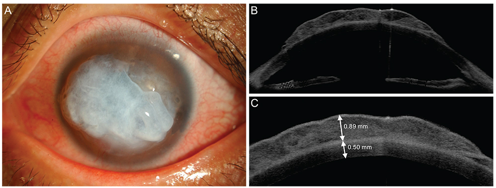
Fig. 1 (A) A preoperative anterior segment photograph of the left eye shows a jelly-like corneal degeneration (8.7 × 7 mm). (B,C) Anterior optical tomography (Visante OCT) shows a 1.39-mm full-thickness cornea and a 0.89-mm-deep area of corneal degeneration.

Fig. 2 (A) A postoperative anterior segment photograph taken 90 days after deep anterior lamellar keratoplasty of the left eye showing a completely healed cornea. (B) Almost corneal epithelial defects had healed by three months after surgery.
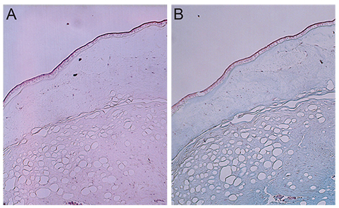
Fig. 3 The lesion is covered by degenerated squamous epithelium. The underlying stroma shows marked myxoid and vacuolar degeneration, with positive Alcian blue staining. A few fibroblast-like spindle cells are scattered in the degenerated stroma. (A) Hematoxylin and eosin, ×40; (B) Alcian blue stain, ×40.

Fig. 4 A high-magnification view of the stroma shows uninucleated (arrowhead) and multinucleated (arrow) spindle cells scattered throughout the myxoid stroma (Alcian blue stain, ×200).
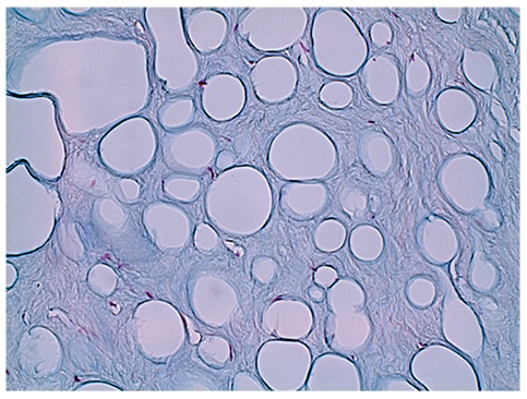
Fig. 5 Marked vacuolar changes in the myxoid corneal stroma (Alcian blue stain, ×200).

Fig. 6 (A) The periphery of the lesion showing a loose, hypocellular, minimal collagen-depositional, glycosaminoglycan-rich lesion in the subepithelial anterior stroma (M), in sharp contrast to the normal deep stroma (N). (B) The central site of the lesion showing vacuolar changes in the deep stroma (V). Masson trichrome stain, ×100.
Reference
-
1. Bulkley BH, Hutchins GM. Atrial myxomas: a fifty year review. Am Heart J. 1979; 97:639–643.2. Horie Y, Ikawa S, Okamoto I, et al. Myxoma of the conjunctiva: a case report and a review of the literature. Jpn J Ophthalmol. 1995; 39:77–82.3. Wollensak G, Green WR, Seiler T. Corneal myxoma. Jpn J Ophthalmol. 2002; 46:193–197.4. Hansen LH, Prause JU, Ehlers N, Heegaard S. Primary corneal myxoma. Acta Ophthalmol Scand. 2004; 82:224–227.5. Chang HJ. Superficial corneal growth. JAMA. 2011; 305:2467–2468.6. Soong T, Soong V, Salvi SM, et al. Primary corneal myxoma. Cornea. 2008; 27:1186–1188.7. Khan AO, Al-Katan H, Al-Gehedan S. Infantile corneal myxoma. J AAPOS. 2008; 12:207–209.8. Robinson JW, Brownstein S, Mintsioulis G. Corneal myxoma arising in a patient with repeated phototherapeutic keratectomies. Cornea. 2006; 25:1111–1114.9. Daoud YJ, Smith R, Smith T, et al. The intraoperative impression and postoperative outcomes of gamma-irradiated corneas in corneal and glaucoma patch surgery. Cornea. 2011; 30:1387–1391.10. Li J, Yu L, Deng Z, et al. Deep anterior lamellar keratoplasty using acellular corneal tissue for prevention of allograft rejection in high-risk corneas. Am J Ophthalmol. 2011; 152:762–770.11. Enzinger FM. Intramuscular myxoma: a review and follow-up study of 34 cases. Am J Clin Pathol. 1965; 43:104–113.12. Perez-Grossmann RA, Mesias LA, Contreras F, Spencer WH. Solitary corneal myxoma. Cornea. 1997; 16:498–500.13. Stout AP. Myxoma, the tumor of primitive mesenchyme. Ann Surg. 1948; 127:706–719.14. Wilbanks GA, Boerner S, Smith D, Rootman DS. Giant pseudocyst formation associated with chronic corneal edema. Cornea. 1997; 16:224–226.15. Margo CE, Mosteller MW. Corneal pseudocyst following acute hydrops. Br J Ophthalmol. 1987; 71:359–360.16. Holden BA, Sweeney DF, Vannas A, et al. Effects of long-term extended contact lens wear on the human cornea. Invest Ophthalmol Vis Sci. 1985; 26:1489–1501.17. Tsubota K, Kaido M, Monden Y, et al. A new surgical technique for deep lamellar keratoplasty with single running suture adjustment. Am J Ophthalmol. 1998; 126:1–8.18. Erickson GA, Landgraf JG, Wessman SJ, et al. Detection and elimination of adventitious agents in continuous cell lines. Dev Biol Stand. 1989; 70:59–66.19. House C, House JA, Yedloutschnig RJ. Inactivation of viral agents in bovine serum by gamma irradiation. Can J Microbiol. 1990; 36:737–740.20. Miekka SI, Forng RY, Rohwer RG, et al. Inactivation of viral and prion pathogens by gamma-irradiation under conditions that maintain the integrity of human albumin. Vox Sang. 2003; 84:36–44.21. Shimazaki J, Yang HY, Tsubota K. Amniotic membrane transplantation for ocular surface reconstruction in patients with chemical and thermal burns. Ophthalmology. 1997; 104:2068–2076.22. Kim JS, Kim JC, Hahn TW, Park WC. Amniotic membrane transplantation in infectious corneal ulcer. Cornea. 2001; 20:720–726.
- Full Text Links
-
- Actions
-
Cited
- CITED
-
- Close
- Share
-
- Similar articles
-
- A Case of Tectonic Lamellar Corneal Patch Graft Using Acellular Cornea in Corneal Ulcer Perforation
- Lamellar Graft of an Acellular, Preserved Human Cornea for Recurrent Anterior Granuloma in Stevens-Johnson Syndrome
- Intramuscular Myxoma of the Foot: A Case Report
- A Case of Conjunctival Myxoma Invading the Caruncle
- Recurrent Myxoma of Maxilla
