A Case of Tectonic Lamellar Corneal Patch Graft Using Acellular Cornea in Corneal Ulcer Perforation
- Affiliations
-
- 1Department of Ophthalmology, Dongguk University Gyeongju Hospital, Dongguk University College of Medicine, Gyeongju, Korea. suksu@dongguk.ac.kr
- KMID: 2215083
- DOI: http://doi.org/10.3341/jkos.2015.56.8.1278
Abstract
- PURPOSE
We report a case of tectonic lamellar corneal patch graft using acellular corneal tissue (Halo Sterile Cornea; Lions VisionGift, Portland, OR, USA) for treating a large corneal ulcer perforation
CASE SUMMARY
A 72-year-old male previously treated for corneal ulcer was referred after presenting with decreased vision and abrupt tears in the right eye. His best-corrected visual acuity was 0.025 (20/800) and slit-lamp examination showed collapsed anterior chamber and 2 x 2 mm corneal perforation with protruded iris at the peripheral cornea. Infiltration in superficial stroma was observed near the perforation. Despite conjunctival flap, the cornea showed leakage due to perforation. To preserve ocular integrity, the patient underwent tectonic lamellar corneal patch graft using acellular corneal tissue. After surgery, the corneal patch graft was well attached. Re-epithelialization occurred after 3 days. There was no recurrence of perforation or corneal graft melting. Visual acuity improved to 0.32 (20/63) after 6 months.
CONCLUSIONS
Tectonic lamellar corneal patch graft using acellular corneal tissue can be a useful treatment option in large corneal ulcer perforation located at the periphery.
MeSH Terms
Figure
-
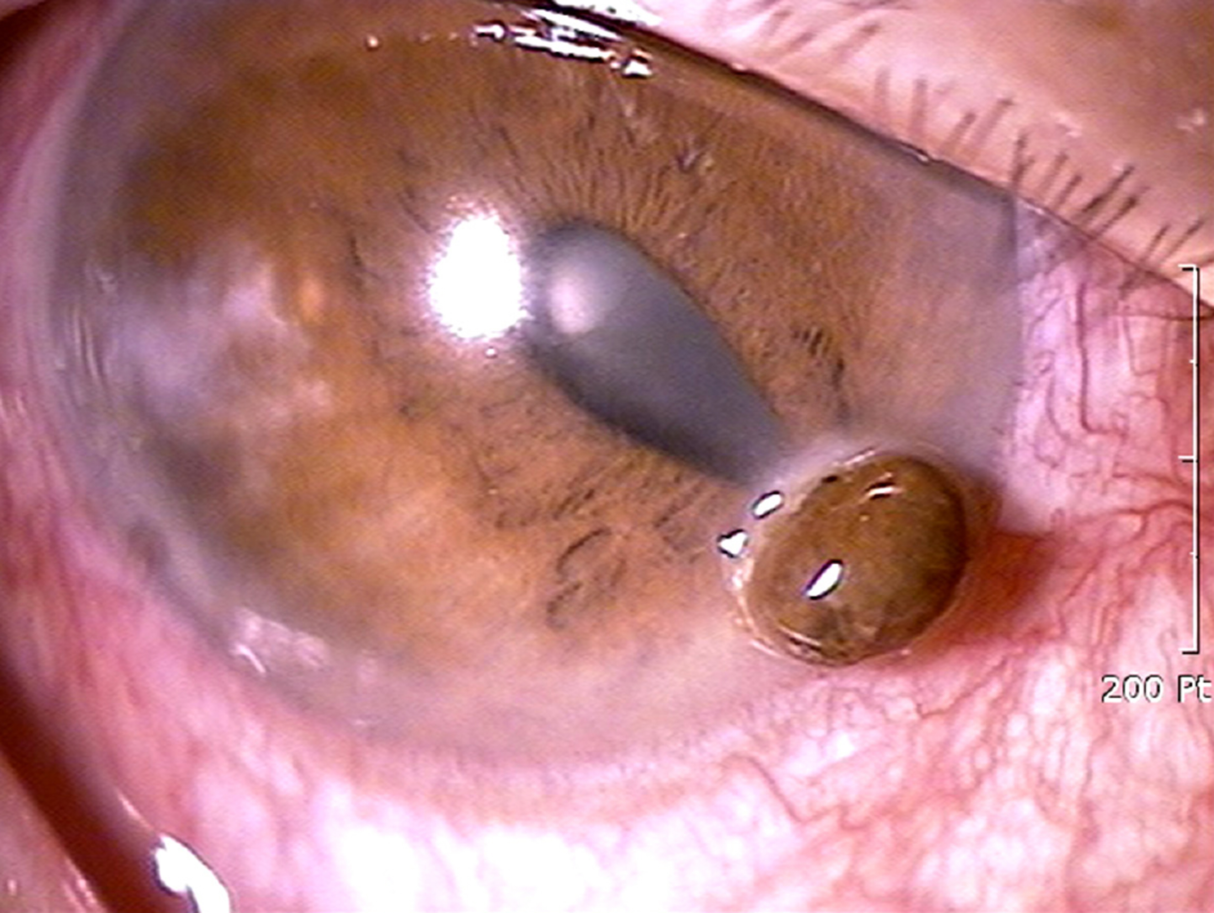
Figure 1. Anterior segment photograph at the initial presentation. The slit-lamp examination reveals large peripheral corneal ul-cer perforation with collapsed anterior chamber. Central ante-rior chamber depth was measured of 2 corneal thickness.

Figure 2. Anterior segment photograph after conjunctival flap. The next day, after conjunctival flap, the slit-lamp ex-amination reveals wound leakage and collapsed anterior cham-ber, despite conjunctival flap.
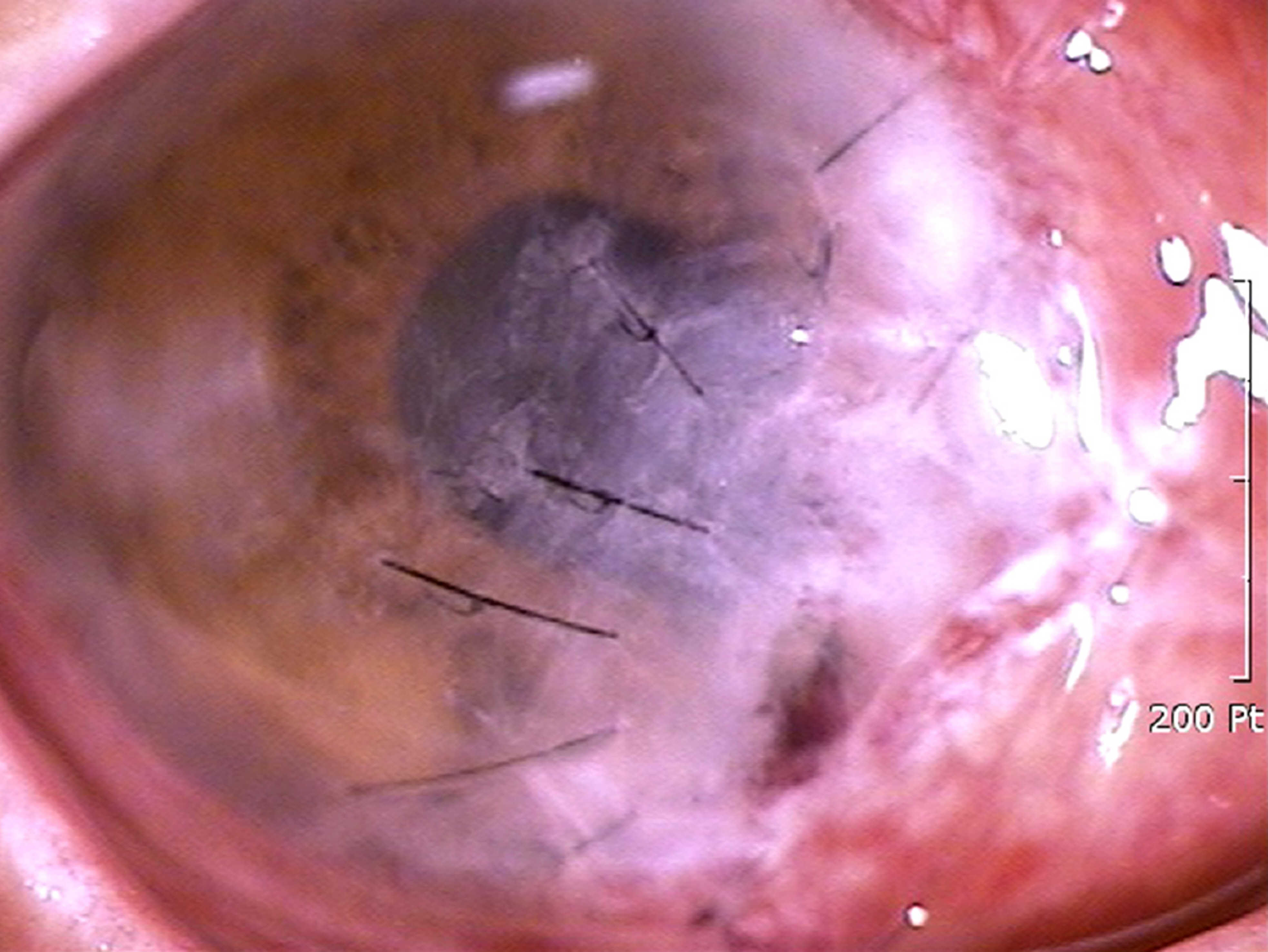
Figure 3. Anterior segment photograph at POD 1. The next day, after tectonic lamellar patch graft, the slit-lamp examina-tion reveals well attached corneal patch graft without leakage. POD = postoperative day.

Figure 4. Anterior segment photograph at POD 3. Three days after tectonic lamellar patch graft, the graft was re-epi-thelialized with normal anterior chamber depth and BCVA of 0.08 (20/250), due to irregular astigmatism. POD = post-operative day; BCVA = best corrected visual acuity.

Figure 5. Anterior segment photograph at POD 1 month. 30 days after patch graft, the slit-lamp examination revealed clear graft with normal anterior chamber depth and BCVA of 0.1 (20/200). POD = postoperative day; BCVA = best corrected visual acuity.

Figure 6. Anterior segment photograph at POD 6 month. After 6 months, the cornea maintained its contour and struc-ture without any complications, with BCVA of 0.32 (20/63). POD = postoperative day; BCVA = best corrected visual acuity.

Figure 7. Topography and dual Scheimpflug image at POD 6 month. The anterior axial curvature map showed asymmetric bowtie pattern which was superonasally steep (the oppsite site of patch graft). The corneal pachymetry map showed relatively even corneal thickness. The elevation map shows slightly elevated shape at the patch graft site. POD = postoperative day.
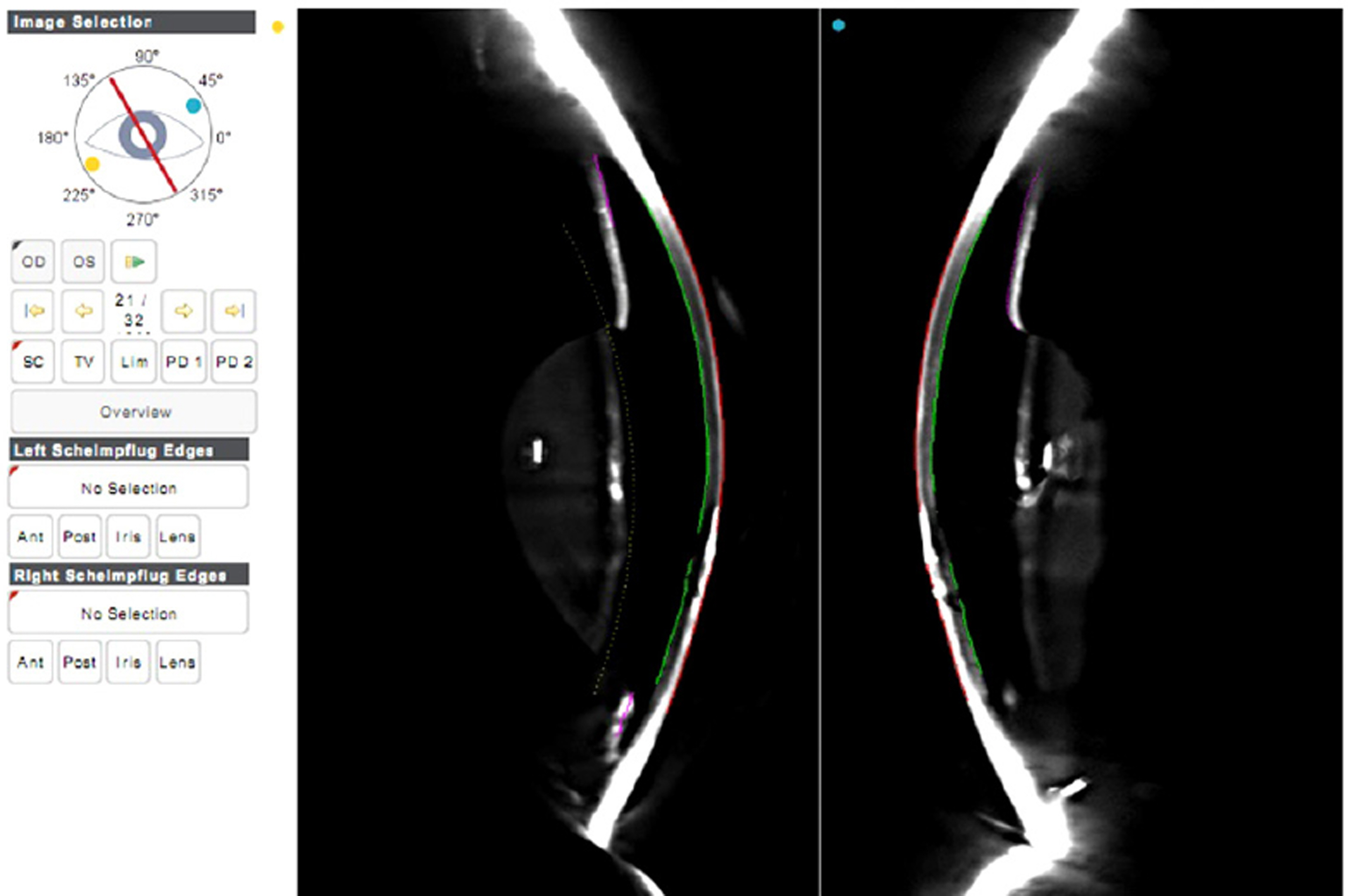
Figure 8. Dual Scheimpflug image of the cornea at POD 6 months. The dual Scheimpflug image of the previously perfo-rated area, shows well attached graft, and relatively normal contour of the anterior and posterior corneal surface. POD = postoperative day.
Cited by 1 articles
-
Corneal Collagen Cross-linking for Corneal Ulcer from Moraxella Group
Sang Earn Woo, Si Hyung Lee
J Korean Ophthalmol Soc. 2020;61(2):200-204. doi: 10.3341/jkos.2020.61.2.200.
Reference
-
References
1. Jeoung SM, Im JS, Park D. A case of conjunctival autotransplantation using conjunctival flap of pterygium in treating corneal ulcer perforation. J Korean Ophthalmol Soc. 2008; 49:2006–10.
Article2. Koh JW, Cho HH, Yang SW, Her J. The effects of the cyanoacrylate tissue adhesive mono-therapy for corneal perforation. J Korean Ophthalmol Soc. 2006; 47:1381–6.3. Vote BJ, Elder MJ. Cyanoacrylate glue for corneal perforations: a description of a surgical technique and a review of the literature. Clin Experiment Ophthalmol. 2000; 28:437–42.
Article4. Sharma N, Sachdev R, Jhanji V. . Therapeutic keratoplasty for microbial keratitis. Curr Opin Ophthalmol. 2010; 21:293–300.
Article5. Oh DH, Kwon MS, Kim JC. Five-layered reinforcing amniotic membrane transplantation for treatment of deep corneal ulcer or perforation. J Korean Ophthalmol Soc. 2011; 52:1232–7.
Article6. Ma DH, Wang SF, Su WY, Tsai RJ. Amniotic membrane graft for the management of scleral melting and corneal perforation in re-calcitrant infectious scleral and corneoscleral ulcers. Cornea. 2002; 21:275–83.
Article7. Shi W, Liu M, Gao H. . Penetrating keratoplasty with small-di-ameter and glycerin-cryopreserved grafts for eccentric corneal perforations. Cornea. 2009; 28:631–7.
Article8. Soong HK, Meyer RF, Sugar A. Small, overlapping tectonic kera-toplasty involving graft-host junction of penetrating keratoplasty. Am J Ophthalmol. 2000; 129:465–7.
Article9. Jeong IY, You IC, Park YG, Yoon KC. Effect of tectonic penetrat-ing keratoplasty for impending perforation due to infectious cor-neal ulcer. J Korean Ophthalmol Soc. 2007; 48:883–8.10. Li J, Yu L, Deng Z. . Deep anterior lamellar keratoplasty using acellular corneal tissue for prevention of allograft rejection in high-risk corneas. Am J Ophthalmol. 2011; 152:762–70.
Article11. Li N, Wang X, Wan P. . Tectonic lamellar keratoplasty with acellular corneal stroma in high-risk corneal transplantation. Mol Vis. 2011; 17:1909–17.12. Daoud YJ, Smith R, Smith T. . The intraoperative impression and postoperative outcomes of gamma-irradiated corneas in cor-neal and glaucoma patch surgery. Cornea. 2011; 30:1387–91.
Article13. Soong HK, Farjo AA, Katz D. . Lamellar corneal patch grafts in the management of corneal melting. Cornea. 2000; 19:126–34.
Article14. Vanathi M, Sharma N, Titiyal JS. . Tectonic grafts for corneal thinning and perforations. Cornea. 2002; 21:792–7.
Article15. Titiyal JS, Ray M, Sharma N. . Intralamellar autopatch with la-mellar keratoplasty for paracentral corneal perforations. Cornea. 2002; 21:615–8.
Article
- Full Text Links
-
- Actions
-
Cited
- CITED
-
- Close
- Share
-
- Similar articles
-
- Tectonic Deep Anterior Lamellar Keratoplasty in Impending Corneal Perforation Using Cryopreserved Cornea
- Lamellar Graft of an Acellular, Preserved Human Cornea for Recurrent Anterior Granuloma in Stevens-Johnson Syndrome
- Multilayer Collagen Sheet Graft for Corneal Perforation with a Corneal Ulcer
- Clinical Evaluation of Full-thickness Deep Lamellar Keratoplasty
- Effect of Tectonic Penetrating Keratoplasty for Impending Perforation Due to Infectious Corneal Ulcer
