Locking Compression Plate in Musculoskeletal Oncology 'a Friend in Need'
- Affiliations
-
- 1Section of Orthopaedic Surgery, Department of Orthopaedic Surgery, The Aga Khan University Hospital, Karachi, Pakistan.
- 2Department of Orthopaedic Surgery, Islam Medical and Dental College, Siakot, Pakistan. kashah_pk@yahoo.com
- KMID: 1787036
- DOI: http://doi.org/10.4055/cios.2013.5.4.321
Abstract
- BACKGROUND
We are presenting our experience in the use of locking compression plate (LCP) after juxta-articular oncological resections in addition to its use in pathologic fracture.
METHODS
A retrospective audit of skeletal reconstruction using LCP in 25 cases of long bone tumors was performed from 2008 to 2010. Reconstruction following limb salvage surgery was done in 17 patients and internal fixation of pathological fracture was done in 8 patients. All patients were available for > 12 months of follow-up, and thus assessed for union at the resected ends.
RESULTS
There were 8 males and 17 females in the study. The average age at the time of surgery was 30 years (range, 9 to 66 years). The minimum follow-up was 12 months (range, 12 to 32 months). All patients except three went on to heal successfully. Complications occurred in those three patients: wound infection in one, nonunion in another, and periprosthetic fracture in the other patient. In the remaining patients, union was achieved at an average of 6.5 months after reconstruction in curative resection and 4.75 months after fixation of pathological fractures.
CONCLUSIONS
Joint sparing limb salvage surgery was made successfully possible after sekeletal reconstruction with LCP. Its use was also quite effective in pathological fractures with poor bone quality. Use of locking plates for musculoskeletal oncological reconstruction resulted in a good and predictable rate of union.
MeSH Terms
Figure
-
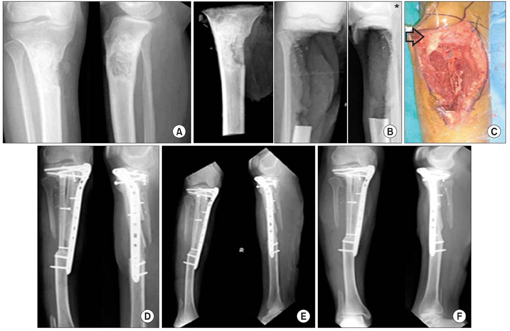
Fig. 1 (A) Preoperative radiograph of osteogenic sarcoma of proximal tibia. (B) Specimen radiographs of the excised bone and residual bone including knee joint. (C) Clinical photograph showing thin slice of remaining proximal tibia along with tibial tuberosity. (D) Postoperative radiograph after reconstruction with vascularised (ipsilateral) and non vascularised fibula. (E) Radiologic union at 6 months. (F) X-rays at 30-month follow-up. *Tibial tuberosity.
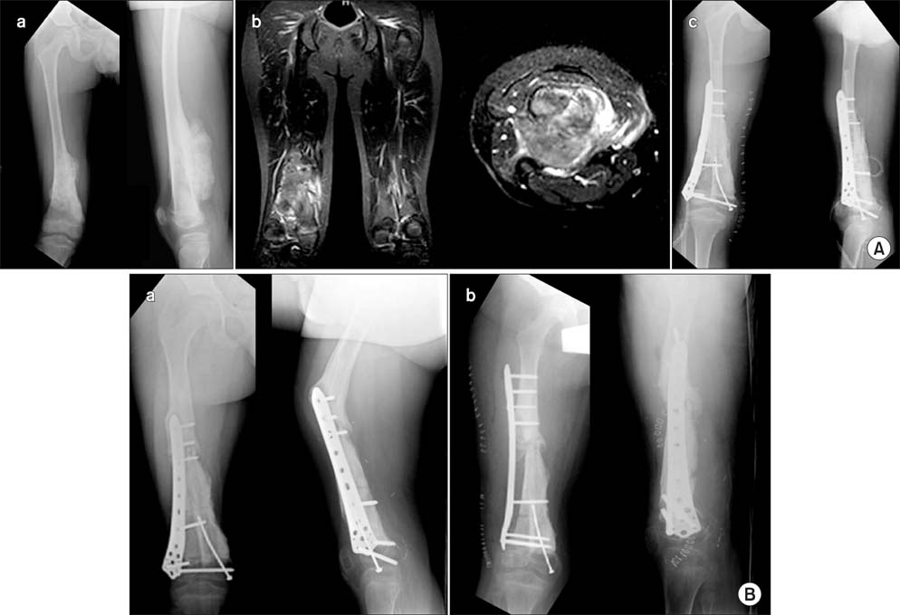
Fig. 2 (A) (a) Preoperative images showing distal femoral lesion (osteogenic sarcoma). (b) Magnetic resonance image showing the extent of lesion and sparing neurovascular bundles. (c) Immediate postoperative X-rays showing reconstruction with autoclaved bone and fibula and osteosynthesis with locking compression plate for distal femoral fracture. (B) (a) Nine-month postoperative X-ray showing angulation in saggital plane at the site of delayed union. (b) Revision of osteosynthesis with a longer plate and correction of angulation.
Reference
-
1. Yu XC, Xu M, Song RX, Xu SF. Marginal resection for osteosarcoma with effective preoperative chemotherapy. Orthop Surg. 2009; 1(3):196–202.2. Schneiderbauer MM, Gullerud R, Harmsen WS, Scully SP. Fibular osteosarcomas: contaminated margins may not impact survival. Clin Orthop Relat Res. 2007; 456:182–187.3. Egol KA, Kubiak EN, Fulkerson E, Kummer FJ, Koval KJ. Biomechanics of locked plates and screws. J Orthop Trauma. 2004; 18(8):488–493.4. Enneking WF, Campanacci DA. Retrieved human allografts: a clinicopathological study. J Bone Joint Surg Am. 2001; 83-A(7):971–986.5. Enneking WF, Mindell ER. Observations on massive retrieved human allografts. J Bone Joint Surg Am. 1991; 73(8):1123–1142.6. Steinau HU, Daigeler A, Langer S, et al. Limb salvage in malignant tumors. Semin Plast Surg. 2010; 24(1):18–33.7. Khattak MJ, Umer M, Haroon-ur-Rasheed , Umar M. Autoclaved tumor bone for reconstruction: an alternative in developing countries. Clin Orthop Relat Res. 2006; 447:138–144.8. Holt GE, Griffin AM, Pintilie M, et al. Fractures following radiotherapy and limb-salvage surgery for lower extremity softtissue sarcomas: a comparison of high-dose and low-dose radiotherapy. J Bone Joint Surg Am. 2005; 87(2):315–319.9. Gainor BJ, Buchert P. Fracture healing in metastatic bone disease. Clin Orthop Relat Res. 1983; (178):297–302.10. Graci C, Maccauro G, Muratori F, Spinelli MS, Rosa MA, Fabbriciani C. Infection following bone tumor resection and reconstruction with tumoral prostheses: a literature review. Int J Immunopathol Pharmacol. 2010; 23(4):1005–1013.11. Egol KA, Kubiak EN, Fulkerson E, Kummer FJ, Koval KJ. Biomechanics of locked plates and screws. J Orthop Trauma. 2004; 18(8):488–493.12. Haidukewych GJ. Innovations in locking plate technology. J Am Acad Orthop Surg. 2004; 12(4):205–212.13. Virkus WW, Miller BJ, Chye PC, Gitelis S. The use of locking plates in orthopaedic oncology reconstructions. Orthopedics. 2008; 31(5):438.14. Wagner M. General principles for the clinical use of the LCP. Injury. 2003; 34:Suppl 2. B31–B42.15. Gitelis S, Cole BJ. The use of allografts in orthopaedic surgery. Instr Course Lect. 2002; 51:507–520.16. Buecker PJ, Berenstein M, Gebhardt MC, Hornicek FJ, Mankin HJ. Locking versus standard plates for allograft fixation after tumour resection in children and adolescents. J Pediatr Orthop. 2006; 26(5):680–685.17. Rastogi S, Kumar A, Khan SA. Do locking plates have a role in orthopaedic oncological reconstruction. Arch Orthop Trauma Surg. 2010; 130(12):1493–1497.
- Full Text Links
-
- Actions
-
Cited
- CITED
-
- Close
- Share
-
- Similar articles
-
- Failure of Removal of Stripped Locking Screw after Locking Compression Plating
- Treatment of Femur Supracondylar Fracture with Locking Compression Plate
- Comparison of Results of Minimally Invasive Plate Osteosynthesis according to Types of Locking Plate in Distal Femoral Fractures
- Treatment of Distal Femur Fracture with Minimally Invasive Locking Compression Plate Osteosynthesis
- Analysis of the Result Treated with Locking Compression Plate-Distal Tibia and Zimmer Periarticular Locking Plate in Distal Tibia Fracture
