Incidental Non-Cardiac Findings of a Coronary Angiography with a 128-Slice Multi-Detector CT Scanner: Should We Only Concentrate on the Heart?
- Affiliations
-
- 1Department of Radiology, Medical School of Thessaly, Mezourlo, Larissa, 41110, Greece. olgalazoura@yahoo.gr
- 2Department of Anatomy, Medical School of Thessaly, Mezourlo, Larissa, 41110, Greece.
- KMID: 1787015
- DOI: http://doi.org/10.3348/kjr.2010.11.1.60
Abstract
OBJECTIVE
To evaluate the spectrum, prevalence, and significance of incidental non-cardiac findings (INCF) in patients referred for a non-invasive coronary angiography using a 128-slice multi-detector CT (MDCT).
MATERIALS AND METHODS
The study subjects included 1,044 patients; 774 males (mean age, 59.9 years) and 270 females (mean age, 63 years), referred for a coronary CT angiography on a 128-slice MDCT scanner. The scans were acquired from the level of the carina to just below the diaphragm. To evaluate INCFs, images were reconstructed with a large field of view (> 300 mm) covering the entire thorax. Images were reviewed in the axial, coronal, and sagittal planes, using the mediastinal, lung, and bone windows. The INCFs were classified as severe, indeterminate, and mild, based on their clinical importance, and as thoracic or abdominal based on their locations.
RESULTS
Incidental non-cardiac findings were detected in 56% of patients (588 of 1,044), including 435 males (mean age, 65.6 years) and 153 females (mean age, 67.9 years). A total of 729 INCFs were observed: 459 (63%) mild (58% thoracic, 43% abdominal), 96 (13%) indeterminate (95% thoracic, 5% abdominal), and 174 (24%) severe (87% thoracic, 13% abdominal). The prevalence of severe INCFs was 15%. Two severe INCFs were histologically verified as lung cancers.
CONCLUSION
The 128-slice MDCT coronary angiography, in addition to cardiac imaging, can provide important information on the pathology of the chest and upper abdomen. The presence of severe INCFs is not rare, especially in the thorax. Therefore, all organs in the scan should be thoroughly evaluated in daily clinical practice.
Keyword
MeSH Terms
Figure
-

Fig. 1 Description of further work-up of patients with indeterminate and severe incidental non-cardiac findings detected by coronary CT angiography.

Fig. 2 Liver cyst (arrow) and ascites (asterisk) incidentally detected in 71-year-old man who was referred for coronary CT angiography for congestive heart failure.

Fig. 3 Incidentally detected hiatus hernias. A. Hiatus hernia (arrow) is depicted in axial plane in 65-year-old man who underwent coronary CT for atypical chest pain. B. Hiatus hernia (arrow) is depicted in sagittal reconstruction of coronary CT angiography in 67-year-old man, also complaining of atypical chest pain.

Fig. 4 Consolidation of right lung discovered in 56-year-old man with cough who underwent coronary CT angiography for atypical chest pain.
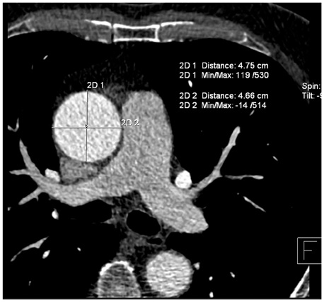
Fig. 5 Aneurysm of ascending aorta measuring 4.7×4.8 cm incidentally found in 62-year-old man who was referred for coronary CT angiography for evaluation of cardiac aetiology of syncope.

Fig. 6 Incidentally detected bronchogenic carcinoma in 65-year-old man. A, B. Mass measuring 4.5 cm in diameter at right hilum is discovered. Pathology revealed squamous cell lung cancer (arrows) on axial plane soft tissue window (A) and coronal plane lung window (B) images.
Reference
-
1. Schoepf UJ, Becker CR, Ohnesorge BM, Yucel EK. CT of coronary artery disease. Radiology. 2004. 232:18–37.2. Achenbach S. Current and future status on cardiac computed tomography imaging for diagnosis and risk stratification. J Nucl Cardiol. 2005. 12:703–713.3. Wann S, Rao P, Des Prez R. Cardiac computed tomographic angiography: evaluation of non-cardiac structures. J Nucl Cardiol. 2009. 16:139–150.4. Kawano Y, Tamura A, Goto Y, Shinozaki K, Zaizen H, Kadota J. Incidental detection of cancers and other non-cardiac abnormalities on coronary multislice computed tomography. Am J Cardiol. 2007. 99:1608–1609.5. Dewey M, Schnapauff D, Teige F, Hamm B. Non-cardiac findings on coronary computed tomography and magnetic resonance imaging. Eur Radiol. 2007. 17:2038–2043.6. Haller S, Kaiser C, Buser P, Bongartz G, Bremerich J. Coronary artery imaging with contrast-enhanced MDCT: extracardiac findings. AJR Am J Roentgenol. 2006. 187:105–110.7. Mueller J, Jeudy J, Poston R, White CS. Cardiac CT angiography after coronary bypass surgery: prevalence of incidental findings. AJR Am J Roentgenol. 2007. 189:414–419.8. Onuma Y, Tanabe K, Nakazawa G, Aoki J, Nakajima H, Ibukuro K, et al. Noncardiac findings in cardiac imaging with multidetector computed tomography. J Am Coll Cardiol. 2006. 48:402–406.9. Gil BN, Ran K, Tamar G, Shmuell F, Eli A. Prevalence of significant noncardiac findings on coronary multidetector computed tomography angiography in asymptomatic patients. J Comput Assist Tomogr. 2007. 31:1–4.10. Law YM, Huang J, Chen K, Cheah FK, Chua T. Prevalence of significant extracoronary findings on multislice CT coronary angiography examinations and coronary artery calcium scoring examinations. J Med Imaging Radiat Oncol. 2008. 52:49–56.11. Budoff MJ, Gopal A. Incidental findings on cardiac computed tomography. Should we look? J Cardiovasc Comput Tomogr. 2007. 1:97–105.12. Hendel RC, Patel MR, Kramer CM, Poon M, Hendel RC, Carr JC, et al. ACCF/ACR/SCCT/SCMR/ASNC/NASCI/SCAI/SIR 2006 appropriateness criteria for cardiac computed tomography and cardiac magnetic resonance imaging: a report of the American College of Cardiology Foundation Quality Strategic Directions Committee Appropriateness Criteria Working Group, American College of Radiology, Society of Cardiovascular Computed Tomography, Society for Cardiovascular Magnetic Resonance, American Society of Nuclear Cardiology, North American Society for Cardiac Imaging, Society for Cardiovascular Angiography and Interventions, and Society of Interventional Radiology. J Am Coll Cardiol. 2006. 48:1475–1497.13. Budoff MJ, Achenbach S, Blumenthal RS, Carr JJ, Goldin JG, Greenland P, et al. Assessment of coronary artery disease by cardiac computed tomography: a scientific statement from the American Heart Association Committee on Cardiovascular Imaging and Intervention, Council on Cardiovascular Radiology and Intervention, and Committee on Cardiac Imaging, Council on Clinical Cardiology. Circulation. 2006. 114:1761–1791.14. Diamond GA, Forrester JS. Analysis of probability as an aid in the clinical diagnosis of coronary-artery disease. N Engl J Med. 1979. 300:1350–1358.15. Kirsch J, Araoz PA, Steinberg FB, Fletcher JG, McCollough CH, Williamson EE. Prevalence and significance of incidental extracardiac findings at 64-multidetector coronary CTA. J Thorac Imaging. 2007. 22:330–334.16. MacMahon H, Austin JH, Gamsu G, Herold CJ, Jett JR, Naidich DP, et al. Guidelines for management of small pulmonary nodules detected on CT scans: a statement from the Fleischner Society. Radiology. 2005. 237:395–400.17. Horton KM, Post WS, Blumenthal RS, Fishman EK. Prevalence of significant noncardiac findings on electron-beam computed tomography coronary artery calcium screening examinations. Circulation. 2002. 106:532–534.18. Hunold P, Schmermund A, Seibel RM, Gronemeyer DH, Erbel R. Prevalence and clinical significance of accidental findings in electron-beam tomographic scans for coronary artery calcification. Eur Heart J. 2001. 22:1748–1758.19. Schragin JG, Weissfeld JL, Edmundowicz D, Strollo DC, Fuhrman CR. Non-cardiac findings on coronary electron beam computed tomography scanning. J Thorac Imaging. 2004. 19:82–86.20. McKenna D, Laxpati M, Colletti PM. The prevalence of incidental findings at cardiac MRI. Open Cardiovasc Med J. 2008. 2:20–25.21. Aglan I, Jodocy D, Hiehs S, Soegner P, Frank R, Haberfellner B, et al. Clinical relevance and scope of accidental extracoronary findings in coronary computed tomography angiography: a cardiac versus thoracic FOV study. Eur J Radiol. 2009. [Epub ahead of print].22. Northam M, Koonce J, Ravenel JG. Pulmonary nodules detected at cardiac CT: comparison of images in limited and full fields of view. AJR Am J Roentgenol. 2008. 191:878–881.23. Yiginer O, Bas S, Pocan S, Yildiz A, Alibek S. Incidental findings of cardiac MSCT: who might benefit from scanning the entire thorax on Ca score imaging? Int J Cardiol. 2008. [Epub ahead of print].24. Kim JW, Kang EY, Yong HS, Kim YK, Woo OH, Oh YW, et al. Incidental extracardiac findings at cardiac CT angiography: comparison of prevalence and clinical significance between precontrast low-dose whole thoracic scan and postcontrast retrospective ECG-gated cardiac scan. Int J Cardiovasc Imaging. 2009. 25:75–81.25. Bach PB, Jett JR, Pastorino U, Tockman MS, Swensen SJ, Begg CB. Computed tomography screening and lung cancer outcomes. JAMA. 2007. 297:953–961.26. Colletti PM. Incidental findings on cardiac imaging. AJR Am J Roentgenol. 2008. 191:882–884.
- Full Text Links
-
- Actions
-
Cited
- CITED
-
- Close
- Share
-
- Similar articles
-
- Diagnostic accuracy of 64-slice multi-detector CT coronary angiography in the evaluation of coronary artery disease
- 16-Slice Multi-Detector Row CT Coronary Angiography: Image Quality and Optimization of the Image Reconstruction Window
- The Diagnostic Accuracy of the 64-slice Multi-detector CT Coronary Angiography for the Assessment of Coronary Artery Stenosis in Symptomatic Patients
- Unusual Coronary Artery Fistula: Left Anterior Descending Coronary Artery - Left Ventricular Fistula Diagnosed by ECG-Gated Multi-Detector Row Coronary CT Angiography
- Clinical Applications of Wide-Detector CT Scanners for Cardiothoracic Imaging: An Update
