J Korean Med Sci.
2008 Jun;23(3):529-532. 10.3346/jkms.2008.23.3.529.
Acute Interstitial Pneumonia in Siblings: A Case Report
- Affiliations
-
- 1Department of Pediatrics and Institution of Allergy, Yonsei University College of Medicine, Seoul, Korea. mhsohn@yumc.yonsei.ac.kr
- 2Department of Radiology, Yonsei University College of Medicine, Seoul, Korea.
- 3Department of Pathology, Yonsei University College of Medicine, Seoul, Korea.
- KMID: 1786896
- DOI: http://doi.org/10.3346/jkms.2008.23.3.529
Abstract
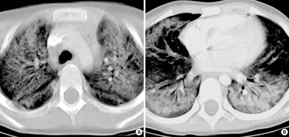
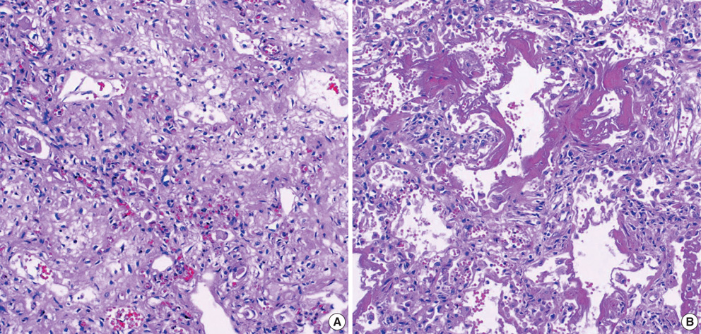
- Acute interstitial pneumonia (AIP) is a rapidly progressive condition of unknown cause that occurs in a previously healthy individual and produces the histologic findings of diffuse alveolar damage. Since the term AIP was first introduced in 1986, there have been very few case reports of AIP in children. Here we present a case of AIP in a 3-yr-old girl whose other two siblings showed similar radiologic findings. The patient was confirmed to have AIP from autopsy showing histological findings of diffuse alveolar damage and proliferation of fibroblasts. Her 3-yr-old brother was also clinically and radiologically highly suspected as having AIP, and the other asymptomatic 8-yr-old sister was radiologically suspected as having AIP.
Keyword
MeSH Terms
Figure
Reference
-
1. Bouros D, Nicholson AC, Polychronopoulos V, du Bois RM. Acute interstitial pneumonia. Eur Respir J. 2000. 15:412–418.
Article2. Wittram C, Mark EJ, McLoud TC. CT-histologic correlation of the ATS/ERS 2002 classification of idiopathic interstitial pneumonias. Radiographics. 2003. 23:1057–1071.
Article3. Katzenstein AL, Myers JL, Mazur MT. Acute interstitial pneumonia. A clinicopathologic, ultrastructural, and cell kinetic study. Am J Surg Pathol. 1986. 10:256–267.4. Vourlekis JS, Brown KK, Cool CD, Young DA, Cherniack RM, King TE, Schwarz MI. Acute interstitial pneumonitis. Case series and review of the literature. Medicine (Baltimore). 2000. 79:369–378.
Article5. Primack SL, Hartman TE, Ikezoe J, Akira M, Sakatani M, Muller NL. Acute interstitial pneumonia: radiographic and CT findings in nine patients. Radiology. 1993. 188:817–820.
Article6. Ichikado K, Johkoh T, Ikezoe J, Takeuchi N, Kohno N, Arisawa J, Nakamura H, Nagareda T, Itoh H, Ando M. Acute interstitial pneumonia: high-resolution CT findings correlated with pathology. AJR Am J Roentgenol. 1997. 168:333–338.
Article7. American Thoracic Society. European Respiratory Society. American Thoracic Society/European Respiratory Society International Multidisciplinary Consensus Classification of the Idiopathic Interstitial Pneumonias. Am J Respir Crit Care Med. 2002. 165:277–304.8. Olson J, Colby TV, Elliott CG. Hamman-Rich syndrome revisited. Mayo Clin Proc. 1990. 65:1538–1548.
Article9. Warner DO, Warner MA, Divertie MB. Open lung biopsy in patients with diffuse pulmonary infiltrates and acute respiratory failure. Am Rev Respir Dis. 1988. 137:90–94.
Article10. Vourlekis JS. Acute interstitial pneumonia. Clin Chest Med. 2004. 25:739–747.
Article
- Full Text Links
- Actions
-
Cited
- CITED
-
- Close
- Share
- Similar articles
-
- Idiopathic Interstitial Pneumonias: Radiologic Findings
- A Case of Nonspecific Interstitial Pneumonia with Clinical Course of Rapid Aggravation
- Effect of interferon-gamma treatment on interstitial pneumonia in a patient with severe combined immunodeficiency
- Idiopathic interstitial pneumonias: clinical findings, pathogenesis, pathology and radiologic findings
- A Case of Asymptomatic, Localized, and Idiopathic Diffuse Alveolar Damage


