The First Korean Case of Cutaneous Lung Tissue Heterotopia
- Affiliations
-
- 1Department of Pediatrics, Inje University College of Medicine, Busan Paik Hospital, Busan, Korea. pedsin@inje.ac.kr
- 2Department of Pathology, Inje University College of Medicine, Busan Paik Hospital, Busan, Korea.
- KMID: 1785924
- DOI: http://doi.org/10.3346/jkms.2010.25.9.1387
Abstract
- Cutaneous lung tissue heterotopia is a very rare disorder where mature lung tissues develop in the skin. This is only the second known report of cutaneous lung tissue heterotopia, with the first by Singer et al. in 1998. A newborn infant had a hemangioma-like, freely movable mass connected to the anterior aspect of the sternal manubrium. Pathologic findings showed mature lung tissues with bronchi, bronchioles, and alveoli through the dermis and subcutis, and it was diagnosed as cutaneous lung tissue heterotopia. Cutaneous lung tissue heterotopia is hypervascular, so grossly it looks like a hemangioma. It can be differentiated from pulmonary sequestration, teratoma, bronchogenic cyst, and branchial cleft cyst by histology and the location of the mass. We describe the clinical, radiologic, and pathologic findings of a cutaneous lung tissue heterotopia, the first reported in Korea.
Keyword
MeSH Terms
Figure
-

Fig. 1 Reddish brown hamangioma-like mass on the sternal manubrium. (A, B) 4.2×3.5×1.0 cm hemangioma-like protruding mass was connected to the middle of sternal manubrium by a stalk. The surface of the mass was wrinkled and vessel-like tissue was visible under the surface. The mass was freely movable and soft.
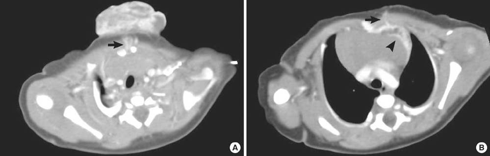
Fig. 2 Chest CT images. (A) Chest CT showed the mass was lobulated, hypervascular, and highly enhanced, and contained two arteries and veins (arrow) that cross the middle of the sternal manubrium. (B) These vessels were connected to the internal mammary arteries and veins (arrow). A similar 1.1×2.3 cm highly enhancing mass (arrowhead) was located in the anterior mediastinum.
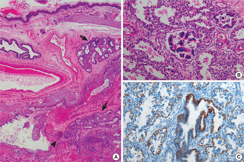
Fig. 3 Histologic findings. (A) Lobulated lung parenchymal tissue (arrow) and a dilated bronchus (arrowhead) with mature cartilages are seen in dermis and subcutis (H&E, ×40). (B) Lung parenchymal tissue is composed of bronchioles lined by columnar epithelia and alveolar spaces containing alveolar macrophages (H&E, ×200). (C) The columnar epithelia and alveolar lining cells are TTF-1 positive.
Cited by 1 articles
-
Pseudomesotheliomatous carcinoma of the lung in the parietal pleura
Ae Ri An, Kyoung Min Kim, Jong Hun Kim, Gong Yong Jin, Young Hoon Choe, Myoung Ja Chung
J Pathol Transl Med. 2020;54(2):192-195. doi: 10.4132/jptm.2019.11.14.
Reference
-
1. Singer G, Haag E, Anabitarte M. Cutaneous lung tissue heterotopia. Histopathology. 1998. 32:60–62.
Article2. Mahler V, Wurm J, Von den Driesch P. Ectopic respiratory epithelium associated with multiple malformations. Br J Dermatol. 1997. 136:933–934.
Article3. Alfadley A, Hainau B, Al Aboud K, Hamadah IR, Al Hawsawi K. Ectopic respiratory mucosa in the skin associated with skeletal malformation and polydactyly. J Am Acad Dermatol. 2000. 43:939–942.
Article4. Conran RM, Stocker JT. Extralobar sequestration with frequently associated congenital cystic adenomatoid malformation, type 2: report of 50 cases. Pediatr Dev Pathol. 1999. 2:454–463.
Article5. Zeidan S, Hery G, Lacroix F, Gorincour G, Potier A, Dubus JC, Guys JM, de Lagausie P. Intralobar sequestration associated with cystic adenomatoid malformation: diagnostic and thoracoscopic pitfalls. Surg Endosc. 2009. 23:1750–1753.
Article6. Tsai TF, Chuan MT, Hsiao CH. A cystic teratoma of the skin. Histopathology. 1996. 29:384–386.
Article7. Moreno A, Muns R. A cystic teratoma in skin. Am J Dermatopathol. 1985. 7:383–386.
Article8. Fraga S, Helwig EB, Rosen SH. Bronchogenic cysts in the skin and subcutaneous tissue. Am J Clin Pathol. 1971. 56:230–238.
Article9. Tresser NJ, Dahms B, Berner JJ. Cutaneous bronchogenic cyst of the back: a case report and review of the literature. Pediatr Pathol. 1994. 14:207–212.
Article10. Jona JZ. Extramediastinal bronchogenic cysts in children. Pediatr Dermatol. 1995. 12:304–306.
Article11. Coleman WR, Homer RS, Kaplan RP. Branchial cleft heterotopia of the lower neck. J Cutan Pathol. 1989. 16:353–358.
Article12. Vure S, Pang K, Hallam L, Lui M, Croaker D. Congenital midline cervical cleft with an underlying bronchogenic like cyst. Pediatr Surg Int. 2009. 25:811–813.
Article
- Full Text Links
-
- Actions
-
Cited
- CITED
-
- Close
- Share
-
- Similar articles
-
- Heterotopic Gastric Mucosa in the Umbilicus
- Nasal Cerebral Heterotopia-so called Nasal Glioma: A case report
- A Case of Neuronal Heterotopia
- A Case of Pseudo-exotropia Caused by Heterotopia of the Macula Associated with Cleft Lip and Palate
- A case of FLNA gene mutation with respiratory insufficiency and periventricular heterotopia
