Curved Periacetabular Osteotomy for the Treatment of Dysplastic Hips
- Affiliations
-
- 1Department of Orthopaedic Surgery, Fukuoka University School of Medicine, Fukuoka, Japan. mnaito@cis.fukuoka-u.ac.jp
- KMID: 1784657
- DOI: http://doi.org/10.4055/cios.2014.6.2.127
Abstract
- Curved periacetabular osteotomy (CPO) was developed for the treatment of dysplastic hips in 1995. In CPO, the exposure of osteotomy sites and osteotomy of the ischium are made in the same manner as Bernese periacetabular osteotomy, and iliac and pubic osteotomies are performed in the same manner as rotational acetabular osteotomy. We studied the dynamic instabilities of 25 dysplastic hips before and after CPO using triaxial accelerometry. Overall magnitude of acceleration was significantly decreased from 2.30 +/- 0.57 m/sec2 preoperatively to 1.55 +/- 0.31 m/sec2 postoperatively. Pain relief and improvement of acetabular coverage resulting from acetabular reorientation seem to be related with reduction of dynamic instabilities of dysplastic hips. Isokinetic muscle strengths of 24 hips in 22 patients were measured preoperatively and after CPO. At 12 months postoperatively, the mean muscle strength exceeded the preoperative values. These results seem to be obtained due to no dissection of abductor muscles in CPO. The preoperative presence of acetabular cysts did not influence the results of CPO. An adequate rotation of the acetabular fragment induced cyst remodeling. Satisfactory results were obtained clinically and radiographically after CPO in patients aged 50 years or older. CPO alone for the treatment of severe dysplastic hips classified as subluxated hips of Severin group IV-b with preoperative CE angles of up to -20degrees could restore the acetabular coverage, weight-bearing area and medialization of the hip joint. CPO without any other combined procedure, as a treatment for 17 hips in 16 patients with Perthes-like deformities, produced good mid-term clinical and radiographic results. We have been performing CPO in conjunction with osteochondroplasty for the treatment of acatabular dysplasia associated with femoroacetabular impingement since 2006. The combined procedure has been providing effective correction of both acetabular dysplasia and associated femoral head-neck deformities without any increased complication rate. We have encountered an obturator artery injury in one case and two intraoperative comminuted fractures. Although serious complications such as motor nerve palsy, deep infection, necrosis of the femoral head or acetabulum, and delayed union or nonunion of the ilium were reported, such complications have never occurred in our 700 cases so far.
Keyword
MeSH Terms
Figure
-
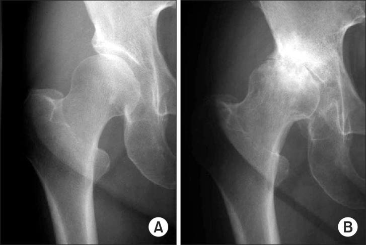
Fig. 1 Radiographs of a 56-year-old female patient with moderate dysplasia of the right hip. (A) Radiograph at presentation showing moderate hip dysplasia with early stage osteoarthritis (OA). (B) Radiograph at 20 months after presentation showing end-stage OA.
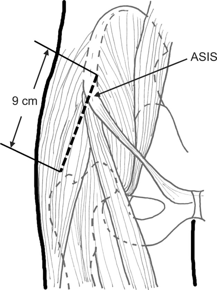
Fig. 2 Schematic drawing of the skin incision. The skin is incised a few centimeters lateral to the course of the lateral femoral cutaneous nerve. ASIS: anterior superior iliac spine.
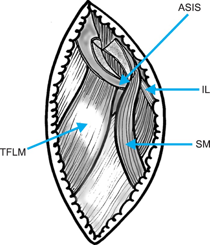
Fig. 3 Schematic drawing of the osteotomy of the anterior superior iliac spine. The anterior superior iliac spine is osteotomized in a wedge-shaped fashion, with the inguinal ligament and sartorius muscle remaining attached in order to prevent the outer table of the pelvis from dissecting. TFLM: tensor fasciae latae muscle, ASIS: anterior superior iliac spine, IL: ilioinguinal ligament, SM: sartorius muscle.
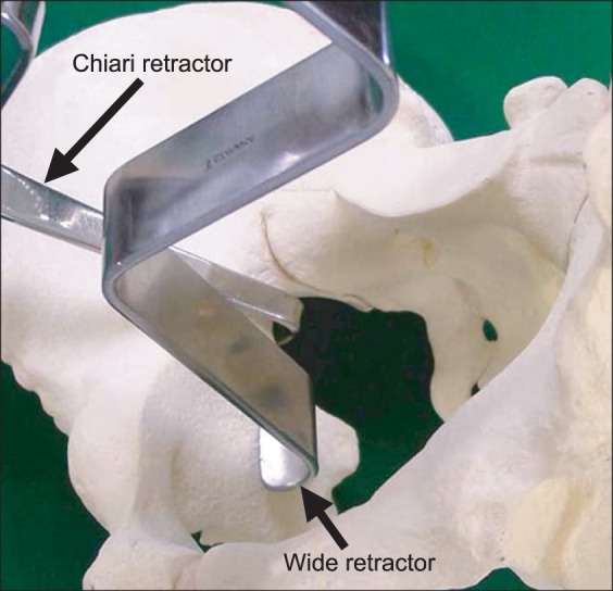
Fig. 4 Photograph of creation of a working space in the quadrilateral space. The blunt tip of a Chiari retractor is fixed in the greater sciatic notch as a landmark. A 3 cm wide retractor is placed in front of the Chiari retractor.
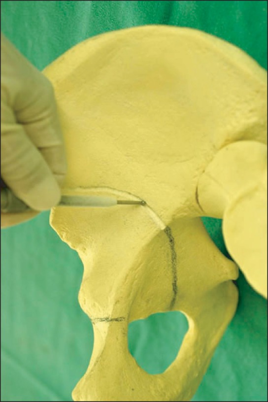
Fig. 5 Photograph of C-shaped osteotomy line of curved periacetabular osteotomy. Using a power drill, a C-shaped osteotomy line is started proximal to the anteroinfer
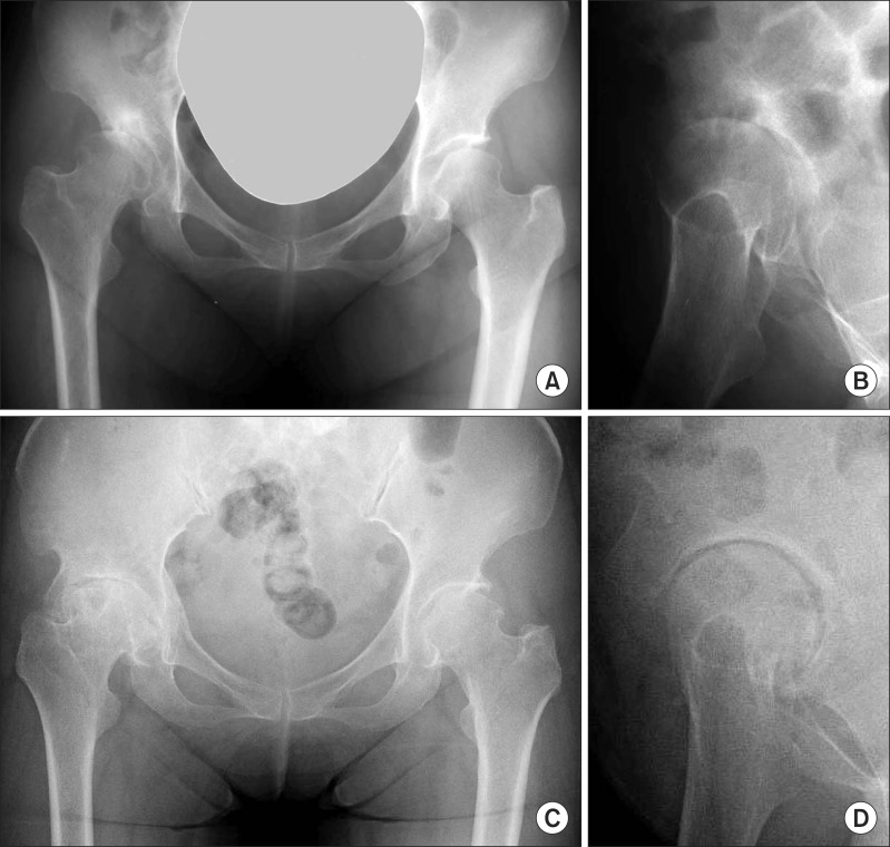
Fig. 6 A 50-year-old woman who underwent curved periacetabular osteotomy (CPO) on her right hip. (A) Preoperative radiograph showing advanced osteoarthritis in her right hip. (B) Preoperative false-profile radiograph showing subluxation of the femoral head in her right hip. (C) Postoperative radiograph at 16 years (age 66), showing improvement of the femoral head coverage in her right hip. The Harris hip score of her right hip improved from 46 points preoperatively to 92 points at 16 years after CPO. (D) Sixteen-year postoperative (age 66) false-profile radiograph showing reduction of the femoral head and good congruity in her right hip. The sourcil of her right hip is markedly enlarged, compared to that before CPO (B).
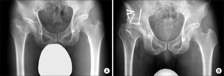
Fig. 7 A 35-year-old man who underwent curved periacetabular osteotomy for the treatment of a dysplastic hip with Perthes-like deformities. (A) Preoperative radiograph showing so-called Perthes-like deformities and retroversion of the acetabulum in his right hip. (B) Five-year postoperative radiograph showing improvement of the femoral head coverage and anteversion of the acetabulum in his right hip. The Harris hip score of his right hip improved from 57 points preoperatively to 98 points postoperatively.
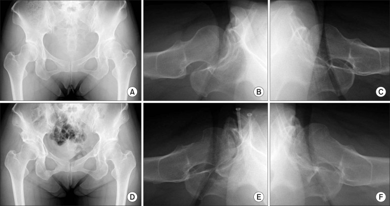
Fig. 8 A 43-year-old woman who underwent curved periacetabular osteotomy in conjunction with osteochondroplasty on both hips. (A) Preoperative radiograph showing moderate acetabular dysplasia in both hips. (B) Preoperative cross-table radiograph showing a slightly prominent head-neck junction of her right hip. (C) Preoperative cross-table radiograph showing a slightly prominent head-neck junction of her left hip. (D) Radiograph made 2 years postsurgery on her left hip (1 year postsurgery on her right hip) showing improvement of the femoral head coverage in both hips. Signs of impingement also disappeared in both hips. (E) Postoperative cross-table radiograph showing recontouring of the femoral head-neck junction of her right hip. The Harris hip score improved from 83 points preoperatively to 94 points at 1 year postsurgery. (F) Postoperative cross-table radiograph showing recontouring of the femoral head-neck junction of her left hip. The Harris hip score improved from 73 points preoperatively to 96 points at 2 years postsurgery.
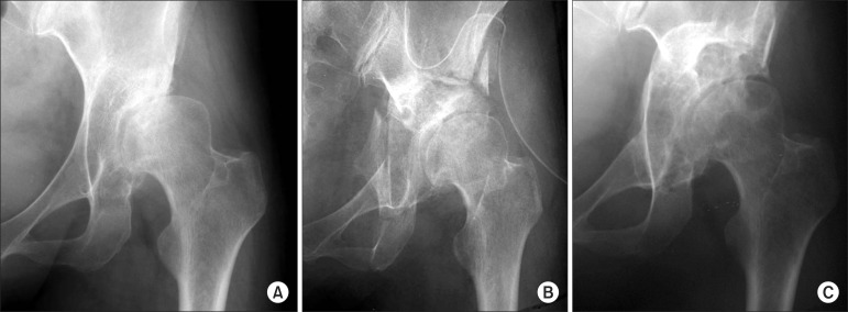
Fig. 9 A 46-year-old woman who had an intraoperative comminuted fracture of the acetabulum during the iliac osteotomy. (A) Preoperative radiograph showing acetabular dysplasia with advanced osteoarthritis in her left hip. (B) Immediate postoperative radiograph showing the comminuted fracture of the osteotomized acetabulum. (C) Radiograph made 6 years postsurgery showing the increased joint space of her left hip. The Harris hip score improved from 61 points preoperatively to 86 points at 6 years postsurgery.
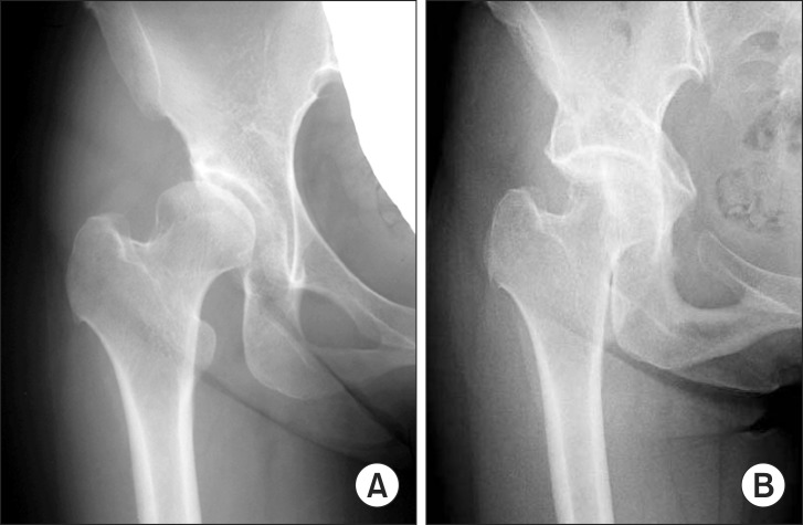
Fig. 10 A 40-year-old-woman who had pubic nonunion after curved periacetabular osteotomy. (A) Preoperative radiograph showing moderate acetabular dysplasia with early stage osteoarthritis in her right hip. (B) Radiograph made 12 years postsurgery showing the pubic nonunion in her right hip. The Harris hip score improved from 96 points preoperatively to 100 points postoperatively.
Reference
-
1. Aronson J. Osteoarthritis of the young adult hip: etiology and treatment. Instr Course Lect. 1986; 35:119–128. PMID: 3819398.2. Nakamura S, Ninomiya S, Nakamura T. Primary osteoarthritis of the hip joint in Japan. Clin Orthop Relat Res. 1989; (241):190–196. PMID: 2924462.
Article3. Takeyama A, Naito M, Shiramizu K, Kiyama T. Prevalence of femoroacetabular impingement in Asian patients with osteoarthritis of the hip. Int Orthop. 2009; 33(5):1229–1232. PMID: 19277653.
Article4. Dorr LD, Kane TJ 3rd, Conaty JP. Long-term results of cemented total hip arthroplasty in patients 45 years old or younger: a 16-year follow-up study. J Arthroplasty. 1994; 9(5):453–456. PMID: 7807101.5. Duffy GP, Berry DJ, Rowland C, Cabanela ME. Primary uncemented total hip arthroplasty in patients <40 years old: 10- to 14-year results using first-generation proximally porous-coated implants. J Arthroplasty. 2001; 16(8 Suppl 1):140–144. PMID: 11742466.6. Burston BJ, Yates PJ, Hook S, Moulder E, Whitley E, Bannister GC. Cemented polished tapered stems in patients less than 50 years of age: a minimum 10-year follow-up. J Arthroplasty. 2010; 25(5):692–699. PMID: 19577886.
Article7. Kim YH, Kim JS, Park JW, Joo JH. Comparison of total hip replacement with and without cement in patients younger than 50 years of age: the results at 18 years. J Bone Joint Surg Br. 2011; 93(4):449–455. PMID: 21464481.8. Sharifi E, Sharifi H, Morshed S, Bozic K, Diab M. Cost-effectiveness analysis of periacetabular osteotomy. J Bone Joint Surg Am. 2008; 90(7):1447–1456. PMID: 18594092.
Article9. Ganz R, Klaue K, Vinh TS, Mast JW. A new periacetabular osteotomy for the treatment of hip dysplasias: technique and preliminary results. Clin Orthop Relat Res. 1988; (232):26–36. PMID: 3383491.
Article10. Millis MB, Murphy SB, Poss R. Osteotomies about the hip for the prevention and treatment of osteoarthrosis. Instr Course Lect. 1996; 45:209–226. PMID: 8727740.
Article11. Naito M, Shiramizu K, Akiyoshi Y, Ezoe M, Nakamura Y. Curved periacetabular osteotomy for treatment of dysplastic hip. Clin Orthop Relat Res. 2005; (433):129–135. PMID: 15805948.
Article12. Ninomiya S, Tagawa H. Rotational acetabular osteotomy for the dysplastic hip. J Bone Joint Surg Am. 1984; 66(3):430–436. PMID: 6699061.
Article13. Maeyama A, Naito M, Moriyama S, Yoshimura I. Evaluation of dynamic instability of the dysplastic hip with use of triaxial accelerometry. J Bone Joint Surg Am. 2008; 90(1):85–92. PMID: 18171961.
Article14. Maeyama A, Naito M, Moriyama S, Yoshimura I. Periacetabular osteotomy reduces the dynamic instability of dysplastic hips. J Bone Joint Surg Br. 2009; 91(11):1438–1442. PMID: 19880886.
Article15. Klaue K, Durnin CW, Ganz R. The acetabular rim syndrome: a clinical presentation of dysplasia of the hip. J Bone Joint Surg Br. 1991; 73(3):423–429. PMID: 1670443.
Article16. Ishiko T, Naito M, Moriyama S. Tensile properties of the human acetabular labrum-the first report. J Orthop Res. 2005; 23(6):1448–1453. PMID: 16099616.
Article17. Kumano T, Naito M, Ishiko T, Moriyama S, Tanaka J. Regional difference of tensile properties of the human acetabular labrum in various hip disorders. Curr Orthop Pract. 2008; 19(5):564–569.
Article18. Xie J, Naito M, Maeyama A. Intracapsular pressure and interleukin-1beta cytokine in hips with acetabular dysplasia. Acta Orthop. 2010; 81(2):189–192. PMID: 20367415.19. Shiramizu K, Naito M, Asayama I, Yatsunami M. A quantitative anatomic characterization of the quadrilateral surface for periacetabular osteotomy. Clin Orthop Relat Res. 2004; (418):157–161. PMID: 15043108.
Article20. Teratani T, Naito M, Shiramizu K, Nakamura Y, Moriyama S. Modified pubic osteotomy for medialization of the femoral head in periacetabular osteotomy: a retrospective study of 144 hips. Acta Orthop. 2008; 79(4):474–482. PMID: 18766479.
Article21. Teratani T, Naito M, Kiyama T, Maeyama A. Periacetabular osteotomy in patients fifty years of age or older: surgical technique. J Bone Joint Surg Am. 2011; 93(Suppl 1):30–39. PMID: 21411684.22. Kuroda D, Maeyama A, Naito M, et al. Dynamic hip stability, strength and pain before and after hip abductor strengthening exercises for patients with dysplastic hips. Isokinet Exerc Sci. 2013; 21(2):95–100.
Article23. Akiyoshi Y, Naito M, Takagishi H, Imai K, Ogata K. Blood flow of the gluteus medius muscle: an animal study. Int Orthop. 1999; 23(4):202–204. PMID: 10591934.24. Ezoe M, Naito M, Asayama I. Muscle strength improves after abductor-sparing periacetabular osteotomy. Clin Orthop Relat Res. 2006; 444:161–168. PMID: 16449917.
Article25. Ezoe M, Naito M, Inoue T. The prevalence of acetabular retroversion among various disorders of the hip. J Bone Joint Surg Am. 2006; 88(2):372–379. PMID: 16452750.
Article26. Kiyama T, Naito M, Shiramizu K, Shinoda T. Postoperative acetabular retroversion causes posterior osteoarthritis of the hip. Int Orthop. 2009; 33(3):625–631. PMID: 18157533.
Article27. Xie J, Naito M, Maeyama A. Evaluation of acetabular versions after a curved periacetabular osteotomy for dysplastic hips. Int Orthop. 2010; 34(4):473–477. PMID: 19424696.
Article28. Ida T, Naito M, Nakamura Y, et al. Improved joint congruency induces joint remodeling after curved periacetabular osteotomy. Hip Joint. 2011; 37:832–835.29. Kinoshita K, Naito M, Nakamura Y, et al. The comparison of old patients after periacetabular osteotomy and younger about joint remodeling. In : AAOS 2011 Annual Meeting; 2011 Feb 16-19; San Diego, CA.30. Nakamura Y, Naito M, Akiyoshi Y, Shitama T. Acetabular cysts heal after successful periacetabular osteotomy. Clin Orthop Relat Res. 2007; 454:120–126. PMID: 16906117.
Article31. Teratani T, Naito M, Kiyama T, Maeyama A. Periacetabular osteotomy in patients fifty years of age or older. J Bone Joint Surg Am. 2010; 92(1):31–41. PMID: 20048093.
Article32. Severin EA. Contribution to the knowledge of congenital dislocation of the hip joint: late results of closed reduction and arthrographic studies of recent cases. Acta Chir Scand. 1941; 84(Suppl 63):1–142.33. Karashima H, Naito M, Shiramizu K, Kiyama T, Maeyama A. A periacetabular osteotomy for the treatment of severe dysplastic hips. Clin Orthop Relat Res. 2011; 469(5):1436–1441. PMID: 20936385.
Article34. Shinoda T, Naito M, Nakamura Y, Kiyama T. Periacetabular osteotomy for the treatment of dysplastic hip with Perthes-like deformities. Int Orthop. 2009; 33(1):71–75. PMID: 17999061.
Article35. Clohisy JC, Nunley RM, Curry MC, Schoenecker PL. Periacetabular osteotomy for the treatment of acetabular dysplasia associated with major aspherical femoral head deformities. J Bone Joint Surg Am. 2007; 89(7):1417–1423. PMID: 17606777.
Article36. Paliobeis CP, Villar RN. The prevalence of dysplasia in femoroacetabular impingement. Hip Int. 2011; 21(2):141–145. PMID: 21484736.
Article37. Clohisy JC, Nunley RM, Carlisle JC, Schoenecker PL. Incidence and characteristics of femoral deformities in the dysplastic hip. Clin Orthop Relat Res. 2009; 467(1):128–134. PMID: 19034600.
Article38. Nassif NA, Schoenecker PL, Thorsness R, Clohisy JC. Periacetabular osteotomy and combined femoral head-neck junction osteochondroplasty: a minimum two-year follow-up cohort study. J Bone Joint Surg Am. 2012; 94(21):1959–1966. PMID: 23138238.39. Nakamura Y, Ida T, Naito M. Clinical results of curved periacetabular osteotomy with osteochondroplasty for acetabular dysplasia with FAI. In : The 86th Annual Meeting of the Japanese Orthopaedic Association; 2013 May 23-26; Hiroshima, Japan.40. Kambe T, Naito M, Asayama I, et al. Vascular anatomy for rotational acetabular osteotomy: cadaveric study. J Orthop Sci. 2003; 8(3):323–328. PMID: 12768473.
Article41. Brenoe AS, Andersen PE, Overgaard S. Endovascular embolisation of severe bleeding in connection with periacetabular osteotomy. Ugeskr Laeger. 2006; 168(14):1453–1454. PMID: 16584678.42. Nishibe N, Tanabe T, Tagawa A, Ninomiya S. A case report of intra pelvic arterial injury in the rotational acetabular osteotomy. Hip Joint. 2008; 28:165–167.43. Kinoshita K, Naito M, Shiramizu K, Kamada S. Prevention of obturator artery injury during pubic osteotomy in periacetabular osteotomy. Curr Orthop Pract. 2011; 22(2):171–175.
Article44. Kamada S, Naito M, Shiramizu K, Nakamura Y, Kinoshita K. Is the obturator artery safe when performing ischial osteotomy during periacetabular osteotomy? Int Orthop. 2011; 35(4):503–506. PMID: 20556381.
Article45. Kiyama T, Naito M, Shiramizu K, Shinoda T, Maeyama A. Ischemia of the lateral femoral cutaneous nerve during periacetabular osteotomy using Smith-Petersen approach. J Orthop Traumatol. 2009; 10(3):123–126. PMID: 19551340.
Article46. Espinosa N, Strassberg J, Belzile EL, Millis MB, Kim YJ. Extraarticular fractures after periacetabular osteotomy. Clin Orthop Relat Res. 2008; 466(7):1645–1651. PMID: 18465184.
Article
- Full Text Links
-
- Actions
-
Cited
- CITED
-
- Close
- Share
-
- Similar articles
-
- Periacetabular Osteotomy for Treating Dysplastic Hips That Were Misdiagnosed as Having Acetabular Bone Tumor: Report of Two Cases
- Periacetabular Osteotomy in Hip Dysplasia with Deformed Femoral Head
- Incidence of Acetabular Retroversion in Dysplastic Hip
- Pelvic Osteotomy in Adults
- Bernese Periacetabular Osteotomy Using Dual Approaches for Hip Dysplasias
