Angioarchitecture of Spinal Dural Arteriovenous Fistula - Evaluation with 3D Rotational Angiography
- Affiliations
-
- 1Departments of Radiology and Research Institute of Radiology, University of Ulsan, College of Medicine, Asan Medical Center, Seoul, Korea. dcsuh@amc.seoul.kr
- 2Department of Neurology, University of Ulsan, College of Medicine, Asan Medical Center, Seoul, Korea.
- 3Department of Neurological Surgery, University of Ulsan, College of Medicine, Asan Medical Center, Seoul, Korea.
- KMID: 1783993
- DOI: http://doi.org/10.5469/neuroint.2012.7.1.10
Abstract
- PURPOSE
The complex angioarchitecture of spinal dural arteriovenous fistulas (SDAVFs) sometimes preclude angiographic analyses or superselective procedures. Therefore, the effectiveness of 3 dimensional rotational angiography (3DRA) as a detailed imaging technique for SDAVFs was evaluated.
MATERIALS AND METHODS
Of 57 patients with spinal vascular malformations, recent 13 SDAVF patients underwent 3DRA. The advantage of 3DRA compared to digital subtraction angiography (DSA) in imaging SDAVF was assessed. Angioarchitecture of SDAVF was focused on location, number, and course of feeders and draining vein. Appropriate angled views were also selected to reveal the segmental artery and feeders.
RESULTS
3DRA technique provided additional information for imaging evaluation of SDAVFs compared to DSA; the presence of multiple feeders, including their transdural portions, as well as their courses. The contralaterally angled anterior-oblique-caudal (spider) view showed the radicular feeder by separating the intercostal artery and the dorsal muscular branch. The bottom-to-up (tunnel) view was useful for revealing the location (ventral vs. dorsal) including sharp medial turn of the dural feeder. The dual mode, which displays both vessels and bones, revealed the course of the feeders and the fistula related to the spinal bony column.
CONCLUSION
Because spinal vasculature overlaps in DSA, 3DRA revealed additional information for evaluations of the number and transdural course of fistular feeders in SDAVFs, and it offers working angles to obtain appropriate views.
MeSH Terms
Figure
-
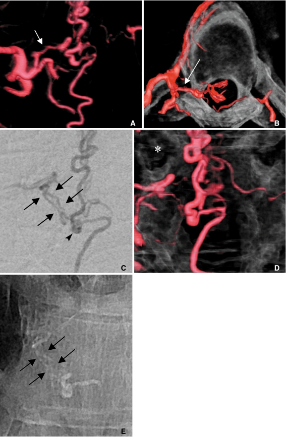
Fig. 1 A 47-year-old male presented with progressive numbness and weakness in both legs.A. The spider view of the right T6 segmental arteriogram shows a radiculodural artery (arrow) running off of the segmental artery.B. The tunnel view shows how acutely the feeder makes a medial turn to enter the intervertebral foramen (a long arrow).C. The selective angiogram shows two parallel channels of the feeder that turn downward toward the fistula (arrows). These two channels can be regarded as the transdural course of the fistula. Note two separate veins ascending and descending directly from the focally dilated fistular end (arrowhead).D. The dual mode (showing vessels and bone) shows how far the fistula is relative to the right pedicle (asterisk) of the T7 vertebra.E. The glue cast penetrated the transdural channels (short arrows) as well as the focally dilated fistular pouch.
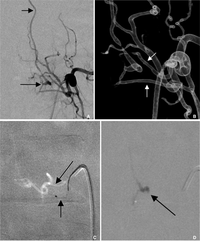
Fig. 2 A 64-year-old female presented with voiding difficulty and weakness in both legs for two months.A. The left L3 lumbar arteriogram shows a SDAVF (arrow) draining into the ascending radicular vein (short arrow).B. The 3D angiogram reveals two separate feeders (arrows) converging onto a fistula.C. The lower feeder (a short arrow) was selected because the upper one (a long arrow) was difficult to select due to the acute angle of its origin.D. A small amount of glue cast penetrated the proximal radicular vein (arrow), occluding the view of the other feeder.
Cited by 2 articles
-
Reversible Symptom Aggravation by Intake of Taurine-Rich Foods in Patients with Venous Congestive Myelopathy: Controlled Case Series Study
Dae Chul Suh, Soo Jeong, Yun Hyeok Choi, Su Min Cho, Su Young Yun, A Yeun Son, Young Min Lim, Boseong Kwon, Yunsun Song
Neurointervention. 2022;17(2):93-99. doi: 10.5469/neuroint.2022.00129.Venous Congestive Myelopathy Caused by Spinal Vascular Malformation
Dae Chul Suh
Neurointervention. 2023;18(2):77-79. doi: 10.5469/neuroint.2023.00262.
Reference
-
1. Thron A, Caplan LR. Brandt T, Caplan LR, Dichgans J, Diener HC, Kennard C, editors. Vascular malformations and interventional neuroradiology of the spinal cord. Neurological disorders: course and treatment. 2003. Amsterdam: Academic Press;p. 517–528.
Article2. Krings T, Geibprasert S. Spinal dural arteriovenous fistulas. AJNR Am J Neuroradiol. 2009; 30:639–648. PMID: 19213818.
Article3. Lee C-S, Pyun HW, Chae EY, Kim K-K, Rhim SC, Suh DC. Reversible aggravation of neurological deficits after steroid medication in patients with venous congestive myelopathy caused by spinal arteriovenous malformation. Interv Neuroradiol. 2009; 15:325–329. PMID: 20465916.
Article4. Jellema K, Tijssen CC, Sluzewski M, van Asbeck FW, Koudstaal PJ, van Gijn J. Spinal dural arteriovenous fistulas-an underdiagnosed disease. A review of patients admitted to the spinal unit of a rehabilitation center. J Neurol. 2006; 253:159–162. PMID: 16222429.5. Suh DC, Choi CG, Sung KB, Kim KK, Rhim SC. Spinal osseous epidural arteriovenous fistula with multiple small arterial feeders converging to a round fistular nidus as a target of venous approach. AJNR Am J Neuroradiol. 2004; 25:69–73. PMID: 14729531.6. Patsalides A, Knopman J, Santillan A, Tsiouris AJ, Riina H, Gobin YP. Endovascular treatment of spinal arteriovenous lesions: beyond the dural fistula. AJNR Am J Neuroradiol. 2011; 32:798–808. PMID: 20651018.
Article7. Jiang L, Huang CG, Liu P, Yan B, Chen JX, Chen HR, et al. 3-Dimensional rotational angiography for the treatment of spinal cord vascular malformations. Surg Neurol. 2008; 69:369–373. PMID: 18262234.
Article8. Prestigiacomo CJ, Niimi Y, Setton A, Berenstein A. Three-dimensional rotational spinal angiography in the evaluation and treatment of vascular malformations. AJNR Am J Neuroradiol. 2003; 24:1429–1435. PMID: 12917141.9. Matsubara N, Miyachi S, Izumi T, Ohshima T, Tsurumi A, Hososhima O, et al. Usefulness of three-dimensional digital subtraction angiography in endovascular treatment of a spinal dural arteriovenous fistula. J Neurosurg Spine. 2008; 8:462–467. PMID: 18447693.
Article10. Aadland TD, Thielen KR, Kaufmann TJ, Morris JM, Lanzino G, Kallmes DF, et al. 3D C-arm conebeam CT angiography as an adjunct in the precise anatomic characterization of spinal dural arteriovenous fistulas. AJNR Am J Neuroradiol. 2010; 31:476–480. PMID: 19850761.
Article11. Berenstein A, Lasjaunias P, ter Brugge KG. Berenstein A, Lasjaunias P, ter Brugge KG, editors. Spinal Dural Arteriovenous Fistulae. Surgical Neuroangiography. Clinical and endovascular treatment aspects in adults. 2003. 2nd ed. Heidelberg: Springer;p. 849–872.
Article12. Berenstein A, Lasjaunias P, ter Brugge KG. Berenstein A, Lasjaunias P, Ter Brugge KG, editors. Spinal and spinal cord arteries and veins. Surgical Neuroangiography. Clincal vascular anatomy and variations. 2001. 2nd ed. New York: Springer;p. 73–164.13. Shi HB, Suh DC, Lee HK, Lim SM, Kim DH, Choi CG, et al. Preoperative transarterial embolization of spinal tumor: embolization techniques and results. AJNR Am J Neuroradiol. 1999; 20:2009–2015. PMID: 10588136.
- Full Text Links
-
- Actions
-
Cited
- CITED
-
- Close
- Share
-
- Similar articles
-
- Image fusion technique using flat panel detector rotational angiography for transvenous embolization of intracranial dural arteriovenous fistula
- Endovascular Treatment of Spinal Dural and Epidural Arteriovenous Fistula as Complication of Lumbar Surgery
- Syringomyelia Associated with Spinal Dural Arteriovenous Fistula: Clinical and Radiological Improvement after Embolization
- A Case of Dural Arteriovenous Fistula of the Anterior Condylar Vein
- Myelopathy due to Spinal Dural Arteriovenous Fistula: A Case Report
