Socket preservation using deproteinized horse-derived bone mineral
- Affiliations
-
- 1Department of Periodontology and Dental Research Institute, Seoul National University School of Dentistry, Seoul, Korea. ccpperio@snu.ac.kr
- KMID: 1783563
- DOI: http://doi.org/10.5051/jpis.2010.40.5.227
Abstract
- PURPOSE
The healing process following tooth extraction apparently results in a pronounced resorption of the alveolar ridge. As a result, the width of alveolar ridge is reduced and severe alveolar bone resorption occurs. The purpose of this experiment is to clinically and histologically evaluate the results of using horse-derived bone mineral for socket preservation.
METHODS
The study comprised 4 patients who were scheduled for extraction as a consequence of severe chronic periodontitis or apical lesion. The extraction was followed by socket preservation using horse-derived bone minerals. Clinical parameters included buccal-palatal width, mid-buccal crest height, and mid-palatal crest height. A histologic examination was conducted.
RESULTS
The surgical sites healed uneventfully. The mean ridge width was 7.75 +/- 2.75 mm at baseline and 7.00 +/- 2.45 mm at 6 months. The ridge width exhibited no significant difference between baseline and 6 months. The mean buccal crest height at baseline was 7.5 +/- 5.20 mm, and at 6 months, 3.50 +/- 0.58 mm. The mean palatal crest height at baseline was 7.75 +/- 3.10 mm, and at 6 months, 5.00 +/- 0.82 mm. There were no significant differences between baseline and 6 months regarding buccal and palatal crest heights. The amount of newly formed bone was 9.88 +/- 2.90%, the amount of graft particles was 42.62 +/- 6.57%, and the amount of soft tissue was 47.50 +/- 9.28%.
CONCLUSIONS
Socket preservation using horse-derived bone mineral can effectively maintain ridge dimensions following tooth extraction and can promote new bone formation through osteoconductive activities.
MeSH Terms
Figure
-

Figure 1 Clinical photograph of the socket preservation procedure. (A) Horse-derived bone minerals were placed into the extraction socket. (B) Primary closure was achieved. (C) Six months of healing. (D) Newly formed bone was incorporated with graft particles at reentry.
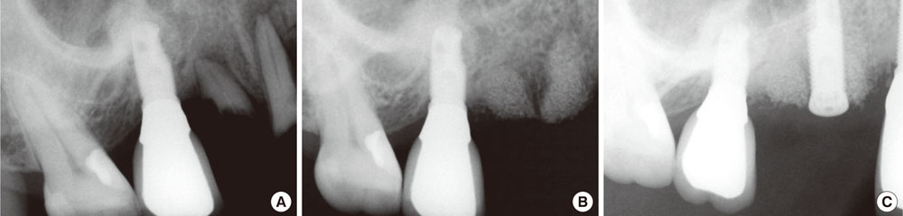
Figure 2 Radiographs of socket preservation and implant placement. (A) Before extraction of retained roots. (B) Six months of healing after placement of horse-derived bone mineral. (C) After implant fixture installation.

Figure 3 Histology from horse-derived bone mineral grafting sites after 6 months. New bones were formed in contact with graft particles (multiple stains; bar = 0.1 mm).
Cited by 1 articles
-
A comparison of different compressive forces on graft materials during alveolar ridge preservation
In-Woo Cho, Jung-Chul Park, Hyun-Seung Shin
J Periodontal Implant Sci. 2017;47(1):51-63. doi: 10.5051/jpis.2017.47.1.51.
Reference
-
1. Schropp L, Wenzel A, Kostopoulos L, Karring T. Bone healing and soft tissue contour changes following single-tooth extraction: a clinical and radiographic 12-month prospective study. Int J Periodontics Restorative Dent. 2003; 23:313–323.2. Araujo MG, Lindhe J. Dimensional ridge alterations following tooth extraction. An experimental study in the dog. J Clin Periodontol. 2005; 32:212–218.
Article3. Tal H. Autogenous masticatory mucosal grafts in extraction socket seal procedures: a comparison between sockets grafted with demineralized freeze-dried bone and deproteinized bovine bone mineral. Clin Oral Implants Res. 1999; 10:289–296.
Article4. Becker W, Urist M, Becker BE, Jackson W, Parry DA, Bartold M, et al. Clinical and histologic observations of sites implanted with intraoral autologous bone grafts or allografts. 15 human case reports. J Periodontol. 1996; 67:1025–1033.
Article5. Froum S, Cho SC, Rosenberg E, Rohrer M, Tarnow D. Histological comparison of healing extraction sockets implanted with bioactive glass or demineralized freeze-dried bone allograft: a pilot study. J Periodontol. 2002; 73:94–102.
Article6. Vance GS, Greenwell H, Miller RL, Hill M, Johnston H, Scheetz JP. Comparison of an allograft in an experimental putty carrier and a bovine-derived xenograft used in ridge preservation: a clinical and histologic study in humans. Int J Oral Maxillofac Implants. 2004; 19:491–497.7. Darby I, Chen ST, Buser D. Ridge preservation techniques for implant therapy. Int J Oral Maxillofac Implants. 2009; 24:Suppl. 260–271.8. Sogal A, Tofe AJ. Risk assessment of bovine spongiform encephalopathy transmission through bone graft material derived from bovine bone used for dental applications. J Periodontol. 1999; 70:1053–1063.
Article9. Amler MH. The time sequence of tissue regeneration in human extraction wounds. Oral Surg Oral Med Oral Pathol. 1969; 27:309–318.
Article10. Botticelli D, Berglundh T, Lindhe J. Hard-tissue alterations following immediate implant placement in extraction sites. J Clin Periodontol. 2004; 31:820–828.
Article11. Araujo MG, Sukekava F, Wennstrom JL, Lindhe J. Ridge alterations following implant placement in fresh extraction sockets: an experimental study in the dog. J Clin Periodontol. 2005; 32:645–652.
Article12. Araujo M, Linder E, Wennstrom J, Lindhe J. The influence of Bio-Oss Collagen on healing of an extraction socket: an experimental study in the dog. Int J Periodontics Restorative Dent. 2008; 28:123–135.13. Pietrokovski J, Massler M. Alveolar ridge resorption following tooth extraction. J Prosthet Dent. 1967; 17:21–27.
Article14. Iasella JM, Greenwell H, Miller RL, Hill M, Drisko C, Bohra AA, et al. Ridge preservation with freeze-dried bone allograft and a collagen membrane compared to extraction alone for implant site development: a clinical and histologic study in humans. J Periodontol. 2003; 74:990–999.
Article15. Artzi Z, Tal H, Dayan D. Porous bovine bone mineral in healing of human extraction sockets. Part 1: histomorphometric evaluations at 9 months. J Periodontol. 2000; 71:1015–1023.
Article16. Lee DW, Pi SH, Lee SK, Kim EC. Comparative histomorphometric analysis of extraction sockets healing implanted with bovine xenografts, irradiated cancellous allografts, and solvent-dehydrated allografts in humans. Int J Oral Maxillofac Implants. 2009; 24:609–615.17. Norton MR, Odell EW, Thompson ID, Cook RJ. Efficacy of bovine bone mineral for alveolar augmentation: a human histologic study. Clin Oral Implants Res. 2003; 14:775–783.
Article18. Carmagnola D, Adriaens P, Berglundh T. Healing of human extraction sockets filled with Bio-Oss. Clin Oral Implants Res. 2003; 14:137–143.
- Full Text Links
-
- Actions
-
Cited
- CITED
-
- Close
- Share
-
- Similar articles
-
- Histopathologic Study of the Effect of two Bovine Bone Powder on Healing of Extraction Socket of Dogs
- Clinical presentation of a horse-derived biomaterial and its Biocompatibility: A Clinical Case Report
- The influence of membrane exposure on post-extraction dimensional change following ridge preservation technique
- Effect of deproteinized bovine bone mineral soaked in inorganic polyphosphate on bone regeneration
- Histologic Analysis of Ridge Preservation Using Deproteinized Porcine Bone: A Retrospective Human Study
