Evaluation of Portal Venous Velocity with Doppler Ultrasound in Patients with Nonalcoholic Fatty Liver Disease
- Affiliations
-
- 1Baskent University Faculty of Medicine Department of Radiology, Adana, Turkey. sulusan@hotmail.com
- 2Baskent University Faculty of Medicine Department of Gastroenterology, Adana Teaching and Medical Research Center, Adana, Turkey.
- KMID: 1783211
- DOI: http://doi.org/10.3348/kjr.2011.12.4.450
Abstract
- PURPOSE
We examined the relationship between portal venous velocity and hepatic-abdominal fat in patients with nonalcoholic fatty liver disease (NAFLD), using spectral Doppler ultrasonography (US) and magnetic resonance imaging (MRI).
MATERIALS AND METHODS
In this prospective study, 35 patients with NAFLD and 29 normal healthy adults (control group) underwent portal Doppler US. The severity of hepatic steatosis in patients with NAFLD was assessed by MRI through chemical shift imaging, using a modification of the Dixon method. Abdominal (intra-abdominal and subcutaneous) fat was measured by MRI.
RESULTS
The difference in portal venous velocity between the patients with NAFLD and the control group was significant (p < 0.0001). There was no correlation between the degree of abdominal or hepatic fat and portal venous velocity (p > 0.05). There were strong correlations between the hepatic fat fraction and subcutaneous adiposity (p < 0.0001), intraperitoneal fat accumulation (p = 0.017), and retroperitoneal fat accumulation (p < 0.0001).
CONCLUSION
Our findings suggest that patients with NAFLD have lower portal venous velocities than normal healthy subjects.
MeSH Terms
-
Abdominal Fat/pathology
Adult
Aged
Blood Flow Velocity/*physiology
Case-Control Studies
Fatty Liver/*physiopathology/*ultrasonography
Female
Humans
Image Interpretation, Computer-Assisted
Magnetic Resonance Imaging
Male
Middle Aged
Obesity/complications
Overweight/complications
Portal Vein/*ultrasonography
Prospective Studies
*Ultrasonography, Doppler
Figure
-

Fig. 1 Spectral Doppler US of maximum velocities in portal veins of control subjects and patients with nonalcoholic steatohepatitis (NAFLD). A. Maximum velocity (Vmax) in portal vein of normal subject (43.30 cm/s). B. Vmax in portal vein of patient with NAFLD (18.10 cm/s).

Fig. 2 In-phase (A) and out-of-phase (B) images, with corresponding signal intensities, are shown for individual with steatosis (32%). Circle in each image represents region of interest. Region of interest signal intensities are from same slice. In-phase and out-of-phase images were used to derive hepatic fat fraction, according to equation: fat fraction = SIin-phase - (SIout-of phase / 2 SIin-phase).
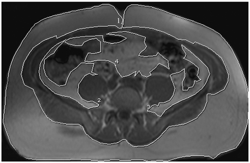
Fig. 3 Axial T1-weighted MR image at umbilical level, with outlined subcutaneous adipose tissue and intra-abdominal adipose tissue (intra- and retro-peritoneal) compartments. Subcutaneous adipose tissue; section 1, retroperitoneal adipose tissue; sections 2 and 3, intraperitoneal adipose tissue; section 4.
Reference
-
1. World Health Organization. Obesity: preventing and managing the global epidemic. Report of a WHO consultation on obesity. WHO/NUT/NCD/981. 1998. Geneva, Switzerland: WHO.2. Wajchenberg BL. Subcutaneous and visceral adipose tissue: their relation to the metabolic syndrome. Endocr Rev. 2000. 21:697–738.3. Mulhall BP, Ong JP, Younossi ZM. Non-alcoholic fatty liver disease: an overview. J Gastroenterol Hepatol. 2002. 17:1136–1143.4. Brunt EM. Pathology of fatty liver disease. Mod Pathol. 2007. 20:Suppl 1. S40–S48.5. Jensen MD, Johnson CM. Contribution of leg and splanchnic free fatty acid (FFA) kinetics to postabsorptive FFA flux in men and women. Metabolism. 1996. 45:662–666.6. Bjorntorp P. "Portal" adipose tissue as a generator of risk factors for cardiovascular disease and diabetes. Arteriosclerosis. 1990. 10:493–496.7. Kissebah AH, Vydelingum N, Murray R, Evans DJ, Hartz AJ, Kalkhoff RK, et al. Relation of body fat distribution to metabolic complications of obesity. J Clin Endocrinol Metab. 1982. 54:254–260.8. Fujioka S, Matsuzawa Y, Tokunaga K, Tarui S. Contribution of intra-abdominal fat accumulation to the impairment of glucose and lipid metabolism in human obesity. Metabolism. 1987. 36:54–59.9. Peiris AN, Struve MF, Mueller RA, Lee MB, Kissebah AH. Glucose metabolism in obesity: influence of body fat distribution. J Clin Endocrinol Metab. 1988. 67:760–767.10. Pilleul F, Chave G, Dumortier J, Scoazec JY, Valette PJ. Fatty infiltration of the liver. Detection and grading using dual T1 gradient echo sequences on clinical MR system. Gastroenterol Clin Biol. 2005. 29:1143–1147.11. Fishbein MH, Mogren C, Gleason T, Stevens WR. Relationship of hepatic steatosis to adipose tissue distribution in pediatric nonalcoholic fatty liver disease. J Pediatr Gastroenterol Nutr. 2006. 42:83–88.12. Fishbein MH, Miner M, Mogren C, Chalekson J. The spectrum of fatty liver in obese children and the relationship of serum aminotransferases to severity of steatosis. J Pediatr Gastroenterol Nutr. 2003. 36:54–61.13. Fishbein MH, Stevens WR. Rapid MRI using a modified Dixon technique: a non-invasive and effective method for detection and monitoring of fatty metamorphosis of the liver. Pediatr Radiol. 2001. 31:806–809.14. Fishbein MH, Gardner KG, Potter CJ, Schmalbrock P, Smith MA. Introduction of fast MR imaging in the assessment of hepatic steatosis. Magn Reson Imaging. 1997. 15:287–293.15. Abate N, Burns D, Peshock RM, Garg A, Grundy SM. Estimation of adipose tissue mass by magnetic resonance imaging: validation against dissection in human cadavers. J Lipid Res. 1994. 35:1490–1496.16. Gronemeyer SA, Steen RG, Kauffman WM, Reddick WE, Glass JO. Fast adipose tissue (FAT) assessment by MRI. Magn Reson Imaging. 2000. 18:815–818.17. Dietrich CF, Lee JH, Gottschalk R, Herrmann G, Sarrazin C, Caspary WF, et al. Hepatic and portal vein flow pattern in correlation with intrahepatic fat deposition and liver histology in patients with chronic hepatitis C. AJR Am J Roentgenol. 1998. 171:437–443.18. Icer S, Kara S. Spectral analysing of portal vein Doppler signals in the cirrhosis patients. Comput Biol Med. 2007. 37:1303–1307.19. Barakat M. Non-pulsatile hepatic and portal vein waveforms in patients with liver cirrhosis: concordant and discordant relationships. Br J Radiol. 2004. 77:547–550.20. Balci A, Karazincir S, Sumbas H, Oter Y, Egilmez E, Inandi T. Effects of diffuse fatty infiltration of the liver on portal vein flow hemodynamics. J Clin Ultrasound. 2008. 36:134–140.21. Garg A. Regional adiposity and insulin resistance. J Clin Endocrinol Metab. 2004. 89:4206–4210.22. Peiris AN, Sothmann MS, Hoffmann RG, Hennes MI, Wilson CR, Gustafson AB, et al. Adiposity, fat distribution, and cardiovascular risk. Ann Intern Med. 1989. 110:867–872.23. Lapidus L, Bengtsson C, Larsson B, Pennert K, Rybo E, Sjostrom L. Distribution of adipose tissue and risk of cardiovascular disease and death: a 12 year follow up of participants in the population study of women in Gothenburg, Sweden. Br Med J (Clin Res Ed). 1984. 289:1257–1261.
- Full Text Links
-
- Actions
-
Cited
- CITED
-
- Close
- Share
-
- Similar articles
-
- Noninvasive serum biomarkers for liver steatosis in nonalcoholic fatty liver disease: Current and future developments
- Nonalcoholic fatty liver disease: pathogenesis and treatment
- Non-invasive Doppler ultrasonography for assessment of the portal hypertension of liver cirrhosis: A prospective study
- The Relation of Nonalcoholic Fatty Liver Disease to Metabolic Syndrome
- Comparison of Doppler Ultrasonography and Hepatic Venous Pressure Gradient in Assessing Portal Hypertension in Liver Cirrhosis
