Segmental Difference of the Hepatic Fibrosis from Chronic Viral Hepatitis due to Hepatitis B versus C Virus Infection: Comparison Using Dual Contrast Material-Enhanced MRI
- Affiliations
-
- 1Department of Radiology and the Research Institute of Radiological Science, Yonsei University College of Medicine, Gangnam Severance Hospital, Seoul 135-720, Korea. yjsrad97@yuhs.ac
- KMID: 1783209
- DOI: http://doi.org/10.3348/kjr.2011.12.4.431
Abstract
OBJECTIVE
We wanted to identify the geographic differences in hepatic fibrosis and their associations with the atrophy-hypertrophy complex in patients with chronic viral hepatitis using the dual-contrast material-enhanced MRI (DC-MRI) with gadopentetate dimeglumine and ferucarbotran.
MATERIALS AND METHODS
Patients with chronic C (n = 22) and B-viral hepatitis (n = 35) were enrolled for determining the subjective grade of fibrosis (the extent and thickness of fibrotic reticulations) in the right lobe (RL), the caudate lobe (CL), the medial segment (MS) and the lateral segment (LS) of the liver, with using a 5-grade scale, on the gradient echo T2*-weighted images of DC-MRI. The fibrosis grades of different segments were compared using the Kruskal-Wallis test followed by post-hoc analysis to establish the segment-by-segment differences. The incidences of two pre-established morphologic signs of cirrhosis were also compared with each other between the two groups of patients.
RESULTS
There were significant intersegmental differences in fibrosis grades of the C-viral group (p = 0.005), and the CL showed lower fibrosis grades as compared with the grades of the RL and MS, whereas all lobes were similarly affected in the B-viral group (p = 0.221). The presence of a right posterior hepatic notch was significantly higher in the patients with intersegmental differences of fibrosis between the RL and the CL (19 out of 25, 76%) than those without such differences (6 out of 32, 19%) (p < 0.001). An expanded gallbladder fossa showed no significant relationship (p = 0.327) with the segmental difference of the fibrosis grades between the LS and the MS.
CONCLUSION
The relative lack of fibrosis in the CL with more advanced fibrosis in the RL can be a distinguishing feature to differentiate chronic C-viral hepatitis from chronic B-viral hepatitis and this is closely related to the presence of a right posterior hepatic notch.
Keyword
MeSH Terms
-
Adult
Aged
Aged, 80 and over
Chi-Square Distribution
Contrast Media/*diagnostic use
Dextrans/*diagnostic use
Diagnosis, Differential
Female
Gadolinium DTPA/*diagnostic use
Hepatitis B, Chronic/*diagnosis
Hepatitis C, Chronic/*diagnosis
Humans
Liver Cirrhosis/*diagnosis/*virology
Magnetite Nanoparticles/*diagnostic use
Male
Middle Aged
Retrospective Studies
Statistics, Nonparametric
Figure
-
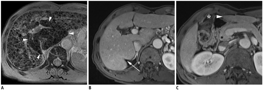
Fig. 1 60-year-old man with C-viral induced cirrhosis. A. Double contrast material-enhanced gradient echo (196/10 msec) T2*-weighted image obtained by 1.5T unit depicts diffuse hyperintense reticulations in entire liver except area of hypertrophic caudate lobe (arrowheads). B. On transverse 3D gradient echo MR images (4.4/2.1 msec) below level of A, right posterior hepatic notch (arrow) is seen between right lobe and caudate lobe. C. Transverse 3D gradient echo MR image (4.4/2.1 msec) demonstrates medial portion of lateral segment (arrowhead) is separated from gallbladder (asterisk) without intervening with medial segment, and this represents expanded gallbladder fossa sign below level of B.
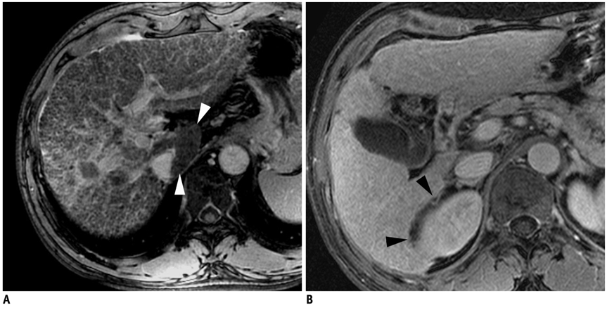
Fig. 2 61-year-old man with C-viral induced cirrhosis. A. Double contrast material-enhanced gradient echo (140/5.8 msec) images obtained by 3T unit depict diffuse involvement of reticular fibrosis involving entire liver except for small portion of caudate lobe (arrowheads). B. There is no hypertrophy of caudate lobe and 3D gradient echo (4.0/1.0 msec) image shows smooth curvature of right posterior medial margin of liver (arrowheads) without any notch formation.
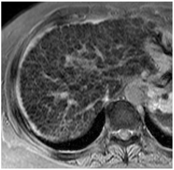
Fig. 3 60-year-old woman with B-viral induced cirrhosis. Double contrast material-enhanced gradient echo (196/10 msec) T2*-weighted hepatic MR image obtained by using 1.5T unit depicts homogeneous reticular fibrosis involving entire liver without any evidence of caudate lobe hypertrophy.
Cited by 2 articles
-
Spectral CT: Preliminary Studies in the Liver Cirrhosis
Peijie Lv, Xiaozhu Lin, Jianbo Gao, Kemin Chen
Korean J Radiol. 2012;13(4):434-442. doi: 10.3348/kjr.2012.13.4.434.Experimental Evaluation of Accelerated T1rho Relaxation Quantification in Human Liver Using Limited Spin-Lock Times
Feng Zhao, Min Deng, Jing Yuan, Gao-Jun Teng, Anil T Ahuja, Yi-Xiang J. Wang
Korean J Radiol. 2012;13(6):736-742. doi: 10.3348/kjr.2012.13.6.736.
Reference
-
1. Custer B, Sullivan SD, Hazlet TK, Iloeje U, Veenstra DL, Kowdley KV. Global epidemiology of hepatitis B virus. J Clin Gastroenterol. 2004. 38:S158–S168.2. Ito K, Mitchell DG. Imaging diagnosis of cirrhosis and chronic hepatitis. Intervirology. 2004. 47:134–143.3. Ito K, Mitchell DG, Gabata T, Hussain SM. Expanded gallbladder fossa: simple MR imaging sign of cirrhosis. Radiology. 1999. 211:723–736.4. Ito K, Mitchell DG, Kim MJ, Awaya H, Koike S, Matsunaga N. Right posterior hepatic notch sign: A simple diagnostic MR finding of cirrhosis. J Magn Reson Imaging. 2003. 18:561–566.5. Okazaki H, Ito K, Fujita T, Koike S, Takano K, Matsunaga N. Discrimination of alcoholic from virus-induced cirrhosis on MR imaging. AJR Am J Roentgenol. 2000. 175:1677–1681.6. Anthony PP, Ishak KG, Nayak NC, Poulsen HE, Scheuer PJ, Sobin LH. The morphology of cirrhosis. Recommendations on definition, nomenclature, and classification by a working group sponsored by the World Health Organization. J Clin Pathol. 1978. 31:395–414.7. Yu JS, Shim JH, Chung JJ, Kim JH, Kim KW. Double contrast-enhanced MRI of viral hepatitis-induced cirrhosis: correlation of gross morphological signs with hepatic fibrosis. Br J Radiol. 2010. 83:212–217.8. Aguirre DA, Behling CA, Alpert E, Hassanein TI, Sirlin CB. Liver fibrosis: noninvasive diagnosis with double contrast material-enhanced MR imaging. Radiology. 2006. 239:425–437.9. Di Lelio A, Cestari C, Lomazzi A, Beretta L. Cirrhosis: diagnosis with sonographic study of the liver surface. Radiology. 1989. 172:389–392.10. Dodd GD 3rd, Baron RL, Oliver JH 3rd, Federle MP. Spectrum of imaging findings of the liver in end-stage cirrhosis: part I, gross morphology and diffuse abnormalities. AJR Am J Roentgenol. 1999. 173:1031–1036.11. Ito K, Mitchell DG, Gabata T. Enlargement of hilar periportal space: a sign of early cirrhosis at MR imaging. J Magn Reson Imaging. 2000. 11:136–140.12. Awaya H, Mitchell DG, Kamishima T, Holland G, Ito K, Matsumoto T. Cirrhosis: modified caudate-right lobe ratio. Radiology. 2002. 224:769–774.13. Castéra L, Vergniol J, Foucher J, Le Bail B, Chanteloup E, Haaser M, et al. Prospective comparison of transient elastography, Fibrotest, APRI, and liver biopsy for the assessment of fibrosis in chronic hepatitis C. Gastroenterology. 2005. 128:343–350.14. Huwart L, Sempoux C, Salameh N, Jamart J, Annet L, Sinkus R, et al. Liver fibrosis: noninvasive assessment with MR elastography versus aspartate aminotransferase-to-platelet ratio index. Radiology. 2007. 245:458–466.15. Zhou X, Lu T, Wei Y, Chen X. Liver volume variation in patients with virus-induced cirrhosis: findings on MDCT. AJR Am J Roentgenol. 2007. 189:w153–w159.16. Starzl TE, Francavilla A, Halgrimson CG, Francavilla FR, Porter KA, Brown TH, et al. The origin, hormonal nature, and action of hepatotrophic substances in portal venous blood. Surg Gynecol Obstet. 1973. 137:179–199.17. Huet PM, Pomier-Layrargues G, Villeneuve JP, Varin F, Viallet A. Intrahepatic circulation in liver disease. Semin Liver Dis. 1986. 6:277–286.18. Popper H. Pathologic aspects of cirrhosis. A review. Am J Pathol. 1977. 87:228–264.19. Shimamatsu K, Kage M, Nakashima O, Kojiro M. Pathomorphological study of HCV antibody-positive liver cirrhosis. J Gastroenterol Hepatol. 1994. 9:624–630.20. Nagula S, Jain D, Groszmann RJ, Garcia-Tsao G. Histological-hemodynamic correlation in cirrhosis-a histological classification of the severity of cirrhosis. J Hepatol. 2006. 44:111–117.21. Ganne-Carrié N, Ziol M, de Ledinghen V, Douvin C, Marcellin P, Castera L, et al. Accuracy of liver stiffness measurement for the diagnosis of cirrhosis in patients with chronic liver diseases. Hepatology. 2006. 44:1511–1517.22. Rosenthal SJ, Harrison LA, Baxter KG, Wetzel LH, Cox GG, Batnitzky S. Doppler US of helical flow in the portal vein. Radiographics. 1995. 15:1103–1111.23. Lafortune M, Matricardi L, Denys A, Favret M, Dery R, Pomier-Layrargues G. Segment 4 (the quadrate lobe): a barometer of cirrhotic liver disease at US. Radiology. 1998. 206:157–160.
- Full Text Links
-
- Actions
-
Cited
- CITED
-
- Close
- Share
-
- Similar articles
-
- Prevention of Viral Hepatitis and Vaccination
- Cytokines in Chronic Hepatitis B and C Virus Infections
- Pre-S Defective Hepatitis B Virus in Patients with Acute and chronic Hepatitis B Virus Infection
- A Case of Coinfection of Hepatitis A and E Virus with Hepatic Encephalopathy
- Management of viral hepatitis in patients with hepatocellular carcinoma
