Infect Chemother.
2012 Feb;44(1):22-25. 10.3947/ic.2012.44.1.22.
Primary Cryptococcal Tenosynovitis in a Patient with Rheumatoid Arthritis
- Affiliations
-
- 1Department of Internal Medicine, Daegu Fatima Hospital, Daegu, Korea. ktkwon@fatima.or.kr
- 2Department of Pathology, Daegu Fatima Hospital, Daegu, Korea.
- KMID: 1782394
- DOI: http://doi.org/10.3947/ic.2012.44.1.22
Abstract
- Here, we report a case of primary cryptococcal tenosynovitis and arthritis caused by worsened cellulitis in a patient with rheumatoid arthritis (RA) who had been taking methotrexate and leflunomide. The patient, injured during the soybean harvest, failed to respond to empirical antibiotic therapy for presumed bacterial cellulitis on the dorsum of the right hand. An operative procedure was performed. Cryptococcocal tenosynovitis was diagnosed upon histopathological examination of the lesion. Treatment with 400 mg of fluconazole daily for 3 months led to the complete disappearance of skin lesions, with slight limitation of finger extension. The patient was examined continuously for 2 years, and there was no evidence of relapse or dissemination to other organs. This case indicates that primary cryptococcal skin and soft tissue infections must be included in the differential diagnoses of antibiotics-refractory soft tissue infections, especially in immunocompromised patients.
Keyword
MeSH Terms
Figure
-

Figure 1 T1 weighted post-gadolinium images (A, B) show a multilobulated and low signaled lesion (arrows) with peripheral soft tissue enhancement along the right fourth and fifth extensor tendons. There is a high signal (arrow) in and around the metacarpophalangeal joint of the right fourth finger in the coronal image (C).
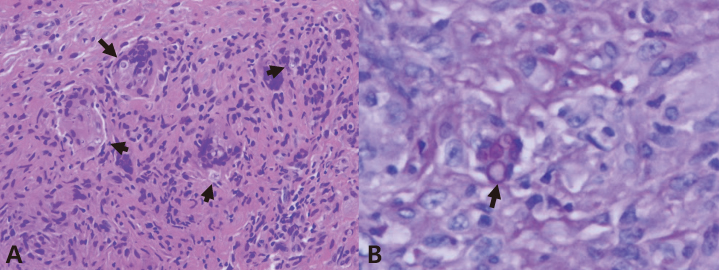
Figure 2 The surgical specimen of the tendon sheath shows granulomatous inflammation with multinucleated giant cells (arrows), which contain small round yeast cells presenting with a foamy cytoplasm surrounded by a clear halo (H & E ×200) (A). Periodic acid-Schiff staining of tissues reveals yeast cells (arrow) that display intense red staining along their rim (PAS ×400) (B).
Reference
-
1. Chayakulkeeree M, Perfect JR. Cryptococcosis. Infect Dis Clin North Am. 2006. 20:507–544. v–vi.
Article2. Lin X, Heitman J. The biology of the Cryptococcus neoformans species complex. Annu Rev Microbiol. 2006. 60:69–105.3. Christianson JC, Engber W, Andes D. Primary cutaneous cryptococcosis in immunocompetent and immunocompromised hosts. Med Mycol. 2003. 41:177–188.
Article4. Neuville S, Dromer F, Morin O, Dupont B, Ronin O, Lortholary O. French Cryptococcosis Study Group. Primary cutaneous cryptococcosis: a distinct clinical entity. Clin Infect Dis. 2003. 36:337–347.
Article5. Hafner C, Linde HJ, Vogt T, Breindl G, Tintelnot K, Koellner K, Landthaler M, Szeimies RM. Primary cutaneous cryptococcosis and secondary antigenemia in a patient with long-term corticosteroid therapy. Infection. 2005. 33:86–89.
Article6. Zorman JV, Zupanc TL, Parac Z, Cucek I. Primary cutaneous cryptococcosis in a renal transplant recipient: case report. Mycoses. 2010. 53:535–537.
Article7. Bruno KM, Farhoomand L, Libman BS, Pappas CN, Landry FJ. Cryptococcal arthritis, tendinitis, tenosynovitis, and carpal tunnel syndrome: report of a case and review of the literature. Arthritis Rheum. 2002. 47:104–108.
Article8. Horcajada JP, Peña JL, Martínez-Taboada VM, Pina T, Belaustegui I, Cano ME, García-Palomo D, Fariñas MC. Invasive Cryptococcosis and adalimumab treatment. Emerg Infect Dis. 2007. 13:953–955.
Article9. Chung SM, Lee EY, Lee CK, Eom DW, Yoo B, Moon HB. A case of cryptococcal tenosynovitis in a patient with Wegener's granulomatosis. J Korean Rheum Assoc. 2004. 11:66–71.10. Park J, Ostrov BE, Schumacher HR Jr. Cryptococcal tenosynovitis in the setting of disseminated cryptococcosis. J Rheumatol. 2000. 27:282–283.11. Kang HY, Kim NS, Lee ES. Primary cutaneous cryptococcosis treated with fluconazole. Korean J Dermatol. 2000. 38:838–840.12. Kim DH, Kim M, Kim SJ, Lee SC, Won YH. A case of primary cutaneous cryptococcosis in a patient with iatrogenic Cushing's syndrome. Korean J Med Mycol. 1998. 3:195–199.13. Kim HJ, Min HG, Lee ES. Two cases of cutaneous cryptococcosis mimicking cellulitis. Korean J Med Mycol. 1998. 3:190–194.14. Kim YJ, Seo SJ, Ro BI. A case of primary cutaneous cryptococcosis misdiagnosel as skin tuberculosis. Korean J Med Mycol. 2003. 8:16–20.15. Probst C, Pongratz G, Capellino S, Szeimies RM, Schölmerich J, Fleck M, Salzberger B, Ehrenstein B. Cryptococcosis mimicking cutaneous cellulitis in a patient suffering from rheumatoid arthritis: a case report. BMC Infect Dis. 2010. 10:239.
Article16. Leão CA, Ferreira-Paim K, Andrade-Silva L, Mora DJ, da Silva PR, Machado AS, Neves PF, Pena GS, Teixeira LS, Silva-Vergara ML. Primary cutaneous cryptococcosis caused by Cryptococcus gattii in an immunocompetent host. Med Mycol. 2011. 49:352–355.
Article17. Pau M, Lallai C, Aste N, Aste N, Atzori L. Primary cutaneous cryptococcosis in an immunocompetent host. Mycoses. 2010. 53:256–258.
Article18. Saag MS, Graybill RJ, Larsen RA, Pappas PG, Perfect JR, Powderly WG, Sobel JD, Dismukes WE. Practice guidelines for the management of cryptococcal disease. Infectious Diseases Society of America. Clin Infect Dis. 2000. 30:710–718.19. Perfect JR, Dismukes WE, Dromer F, Goldman DL, Graybill JR, Hamill RJ, Harrison TS, Larsen RA, Lortholary O, Nguyen MH, Pappas PG, Powderly WG, Singh N, Sobel JD, Sorrell TC. Clinical practice guidelines for the management of cryptococcal disease: 2010 update by the infectious diseases society of america. Clin Infect Dis. 2010. 50:291–322.
Article20. Baumgarten KL, Valentine VG, Garcia-Diaz JB. Primary cutaneous cryptococcosis in a lung transplant recipient. South Med J. 2004. 97:692–695.
Article
- Full Text Links
-
- Actions
-
Cited
- CITED
-
- Close
- Share
-
- Similar articles
-
- Cryptococcal Tenosynovitis of the Hand in a Patient with Rheumatiod Arthritis: A Case Report
- Unusual, but important, peri- and extra-articular manifestations of rheumatoid arthritis: a pictorial essay
- MR Imaging of the Early Rheumatoid Arthritis: Usefulness of Contrast Enhanced Fat Suppressed SPGR Imaging
- A Case of Cryptococcal Tenosynovitis in a Patient with Wegener's Granulomatosis
- A Case of Tenosynovitis Due to Mycobacterium intracellulare in a Patient with Rheumatoid Arthritis
