J Korean Med Sci.
2009 Oct;24(5):956-959. 10.3346/jkms.2009.24.5.956.
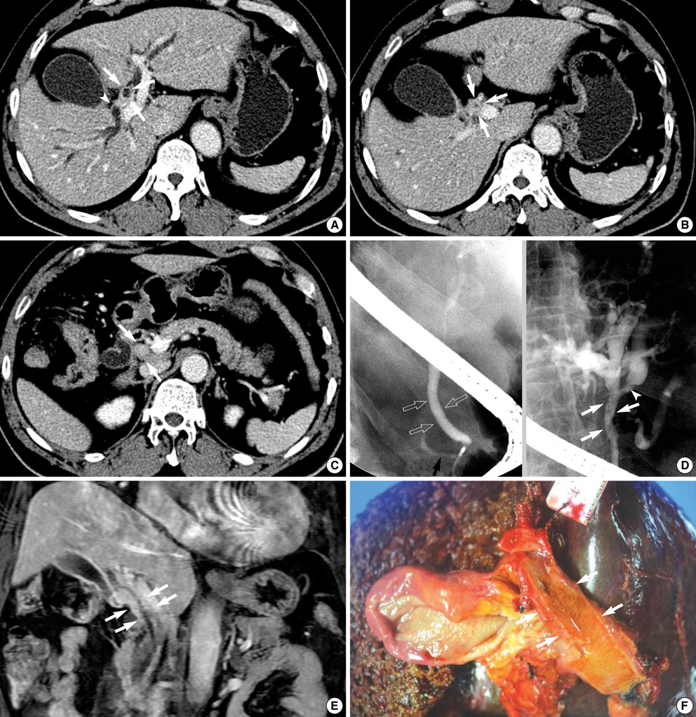
Primary Biliary Lymphoma Mimicking Cholangiocarcinoma: A Characteristic Feature of Discrepant CT and Direct Cholangiography Findings
- Affiliations
-
- 1Department of Radiology, Seoul National University College of Medicine, Seoul, Korea. jm@radcom.snu.ac.kr
- 2Institute of Radiation Medicine, Seoul National University College of Medicine, Seoul, Korea.
- 3Department of Surgery, Seoul National University College of Medicine, Seoul, Korea.
- 4Department of Pathology, Seoul National University College of Medicine, Seoul, Korea.
- KMID: 1782022
- DOI: http://doi.org/10.3346/jkms.2009.24.5.956
Abstract
- Primary non-Hodgkin's lymphoma arising from the bile duct is extremely rare and the reported imaging features do not differ from those of cholangiocarcinoma of the bile duct. We report a case of a patient with extranodal marginal zone B-cell lymphoma of mucosa associated lymphoid tissue (MALT), who presented with obstructive jaundice and describe the distinctive radiologic features that may suggest the correct preoperative diagnosis of primary lymphoma of the bile duct. Primary MALT lymphoma of the extrahepatic bile duct should be considered in the differential diagnosis when there is a mismatch in imaging findings on computed tomography or magnetic resonance imaging and cholangiography.
Keyword
MeSH Terms
-
Bile Duct Neoplasms/complications/*diagnosis/radiography
*Bile Ducts, Extrahepatic
Cholangiocarcinoma/diagnosis
Cholangiography
Diagnosis, Differential
Humans
Jaundice, Obstructive/complications/diagnosis
Lymphoma, B-Cell, Marginal Zone/complications/*diagnosis/radiography
Magnetic Resonance Imaging
Male
Middle Aged
Tomography, X-Ray Computed
Figure
Cited by 1 articles
-
Primary Biliary Mucosa-associated Lymphoid Tissue Lymphoma Mimicking Hilar Cholangiocarcinoma
Seungha Hwang, Tae Jun Song, Seol So, Min Kyung Jeon, Eun Hye Oh, Byoung Soo Kwon, Sujong An, Myung-Hwan Kim
Korean J Gastroenterol. 2016;68(2):114-118. doi: 10.4166/kjg.2016.68.2.114.
Reference
-
1. Kang CS, Lee YS, Kim SM, Kim BK. Primary low grade B cell lymphoma of mucosa associated lymphoid tissue type of the common bile duct. J Gastroenterol Hepatol. 2001. 16:949–951.2. Joo YE, Park CH, Lee WS, Kim HS, Choi SK, Cho CK, Rew JS, Kim SJ, Maetani I. Primary non-Hodgkin's lymphoma of the common bile duct presenting as obstructive jaundice. J Gastroenterol. 2004. 39:692–696.
Article3. Eliason SC, Grosso LE. Primary biliary malignant lymphoma clinically mimicking cholangiocarcinoma: a case report and review of the literature. Ann Diagn Pathol. 2001. 5:25–33.
Article4. Nguyen GK. Primary extranodal non-Hodgkin's lymphoma of the extrahepatic bile ducts. Report of a case. Cancer. 1982. 50:2218–2222.
Article5. Jho DH, Jho DJ, Chejfec G, Ahn M, Ong ES, Espat NJ. Primary biliary B-cell lymphoma of the cystic duct causing obstructive jaundice. Am Surg. 2007. 73:508–510.
Article6. Boccardo J, Khandelwal A, Ye D, Duke BE. Common bile duct MALT lymphoma: case report and review of the literature. Am Surg. 2006. 72:85–88.
Article7. Das K, Fisher A, Wilson DJ, dela Torre AN, Seguel J, Koneru B. Primary non-Hodgkin's lymphoma of the bile ducts mimicking cholangiocarcinoma. Surgery. 2003. 134:496–500.
Article8. Park MS, Kim TK, Kim KW, Park SW, Lee JK, Kim JS, Lee JH, Kim KA, Kim AY, Kim PN, Lee MG, Ha HK. Differentiation of extrahepatic bile duct cholangiocarcinoma from benign stricture: findings at MRCP versus ERCP. Radiology. 2004. 233:234–240.
Article9. Silva MA, Tekin K, Aytekin F, Bramhall SR, Buckels JA, Mirza DF. Surgery for hilar cholangiocarcinoma; a 10 year experience of a tertiary referral centre in the UK. Eur J Surg Oncol. 2005. 31:533–539.
Article
- Full Text Links
- Actions
-
Cited
- CITED
-
- Close
- Share
- Similar articles
-
- Primary Biliary Mucosa-associated Lymphoid Tissue Lymphoma Mimicking Hilar Cholangiocarcinoma
- Retained Intrahepatic Stones' Comparative Study of T-tube Cholangiography, Selective Cholangiography, and Computed Tomography
- Biliary Tract & Pancreas; A Case of Cholangiocarcinoma Suggested as Developing in the Patient with Primary Sclerosing Cholangitis
- Helical CT Cholangiography with Multiplanar Reformation: Utility in Patients with Extrahepatic Biliary Obstruction
- PTBD Spiral CT Cholangiography: Utility in Patients with Extrahepatic Biliary Obstruction


