J Korean Med Sci.
2006 Oct;21(5):950-953. 10.3346/jkms.2006.21.5.950.
Cavernous Angioma in the Falx Cerebri: A Case Report
- Affiliations
-
- 1Department of Neurosurgery, The Catholic University of Korea, Seoul, Korea. hongyk@catholic.ac.kr
- KMID: 1781923
- DOI: http://doi.org/10.3346/jkms.2006.21.5.950
Abstract
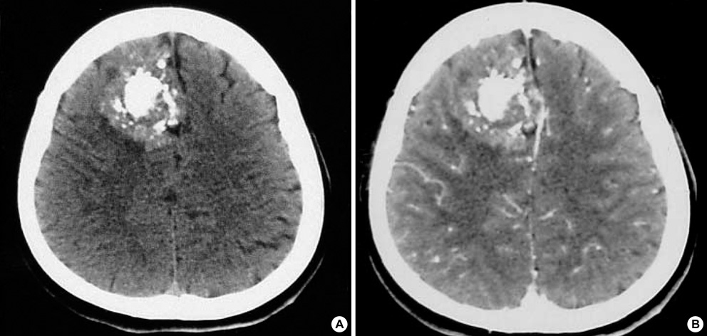
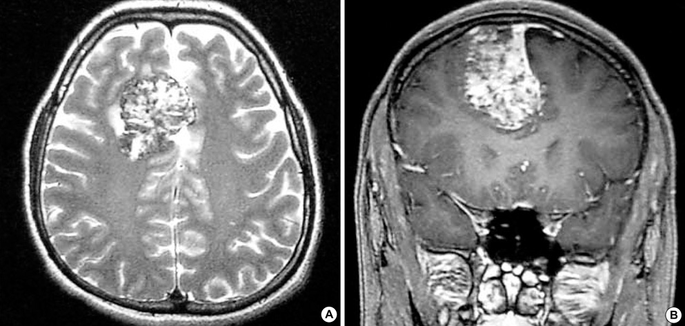
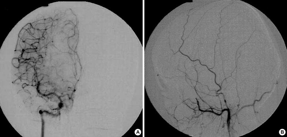
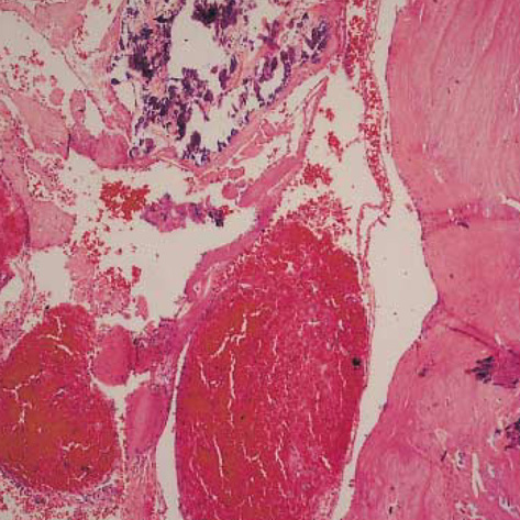
- Intracranial cavernous angiomas are benign vascular malformations and can be divided into intra-axial and extra-axial lesions. Extra-axial cavernous angiomas are relatively rare and usually arise in relation to the dura mater and mimick meningiomas. We report a case of cavernous angioma that occured in the falx cerebri of a 22-yr-old female patient with the special focus on neuroradiologic findings. This is the fourth case of cavernous angioma in the falx cerebri reported in the literature to our knowledge.
MeSH Terms
Figure
Reference
-
1. Curling OD Jr, Kelly DL Jr, Elster AD, Craven TE. An analysis of the natural history of cavernous angiomas. J Neurosurg. 1991. 75:702–708.2. Zabramski JM, Awasthi D. Awad IA, Barrow DL, editors. Extra-axial cavernous malformations. Cavernous malformations. 1993. Park Ridge, IL: American Association of Neurological Surgeons;133–144.3. Biondi A, Clemenceau S, Dormont D, Deladoeuille M, Ricciardi GK, Mokhtari K, Sichez JP, Marsault C. Intracranial extra-axial cavernous (HEM) angiomas: tumors or vascular malformation? J Neuroradiol. 2002. 29:91–104.4. Lee SB, Lee JI, Kim JS, Hong SC, Park K. Treatment of brainstem cavernomas. Korean J Cerebrovasc Surg. 2004. 6:58–63.5. Fracasso L. Considerazioni su un caso di angioma della falce. Riv Patol Nerv Ment. 1947. 68:214–226.6. Kaga A, Isono M, Mori T, Kusakabe T, Okada H, Hori S. Cavernous angioma of falx cerebri; case report. No Shinkei Geka. 1991. 19:1079–1083.7. Ebeling JD, Tranmer BI, Davis KA, Kindt GW, De Masters BK. Thrombosed arteriovenous malformation. Magnetic resonance imaging and histopathological correlations. Neurosurgery. 1988. 23:605–610.8. Robinson JR Jr, Awad IA, Masaryk TJ, Estes ML. Pathological heterogeneity of angiographically occult vascular malformations of the brain. Neurosurgery. 1993. 33:547–555.
Article9. Harper DG, Buck DR, Early CB. Visual loss from cavernous hemangiomas of the middle cranial fossa. Arch Neurol. 1982. 39:252–254.
Article10. Rigamonti D, Drayer BP, Johnson PC, Hadley MN, Zabramski J, Spetzler RF. The MRI appearance of cavernous malformations (angiomas). J Neurosurg. 1987. 67:518–524.
Article11. Sato M, Nakano M, Sasanuma M, Asari J, Watanabe K, Hanai K. MR image of multiple intracranial cavernous angioma: the usefulness of gradient-echo MR image. No To Shinkei. 2003. 55:172–173.12. Guermazi A, Lafitte F, Miaux Y, Adem C, Bonneville JF, Chiras J. The dural tail sign-beyond meningioma. Clin Radiol. 2005. 60:171–188.
Article13. Taniuchi N, Yamazaki M, Katsura K, Igarashi H, Sakamoto S, Katayama Y. T2-weighted MRI in a case with multiple cerebral cavernous malformations. No To Shinkei. 2002. 54:1092–1093.14. Seo Y, Fukuoka S, Sasaki T, Takanashi M, Hojo A, Nakamura H. Cavernous sinus hemangioma treated with gamma knife radiosurgery: usefulness of SPECT for diagnosis--case report. Neurol Med Chir. 2000. 40:575–578.
- Full Text Links
- Actions
-
Cited
- CITED
-
- Close
- Share
- Similar articles
-
- A Case of Oligodendroglioma Attached to the Leptomeninges and Falx Cerebri
- Intra-Root Cavernous Angioma of the Cauda Equina : A Case Report and Review of the Literature
- Cavernous Angioma Coexisting with Venous Angioma in the Posterior Fossa
- A Case of Cavernous Angioma of the Cerebellar Vermis
- Reevaluation of the “falx signâ€


