J Korean Med Sci.
2006 Apr;21(2):365-367. 10.3346/jkms.2006.21.2.365.
A Case of Autoimmune Hemolytic Anemia Associated with an Ovarian Teratoma
- Affiliations
-
- 1Department of Internal Medicine, College of Medicine, Hallym University, Chuncheon, Korea. hunhos@hallym.ac.kr
- 2Department of Obstetrics and Gynecology, College of Medicine, Hallym University, Chuncheon, Korea.
- 3Department of Pathology, College of Medicine, Hallym University, Chuncheon, Korea.
- KMID: 1781853
- DOI: http://doi.org/10.3346/jkms.2006.21.2.365
Abstract
- Autoimmune hemolytic anemia associated with an ovarian teratoma is a very rare disease. However, treating teratoma is the only method to cure the hemolytic anemia, so it is necessary to include ovarian teratoma in the differential diagnosis of autoimmune hemolytic anemia. We report herein on a case of a young adult patient who had severe autoimmune hemolytic anemia that was induced by an ovarian teratoma. A 25-yr-old woman complained of general weakness and dizziness for 1 week. The hemoglobin level was 4.2 g/dL, and the direct and indirect antiglobulin tests were all positive. The abdominal computed tomography scan revealed a huge left ovarian mass, and this indicated a teratoma. She was refractory to corticosteroid therapy; however, after surgical resection of the ovarian mass, the hemoglobin level and the reticulocyte count were gradually normalized. The mass was well encapsulated and contained hair and teeth. She was diagnosed as having autoimmune hemolytic anemia associated with an ovarian teratoma. To the best of our knowledge, this is the first such a case to be reported in Korea.
Keyword
MeSH Terms
Figure
-
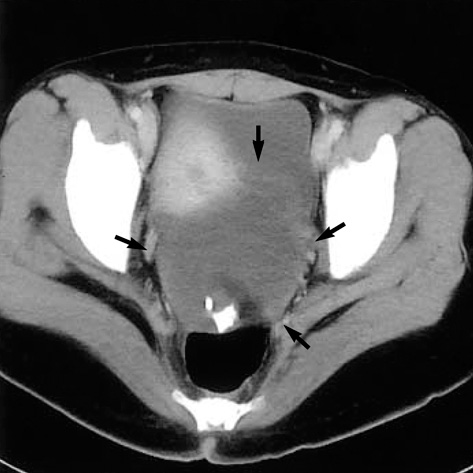
Fig. 1 Abdominal computed tomographic scan shows a huge ovoid mass (arrows) with multiple calcific nodules anterior to rectum.
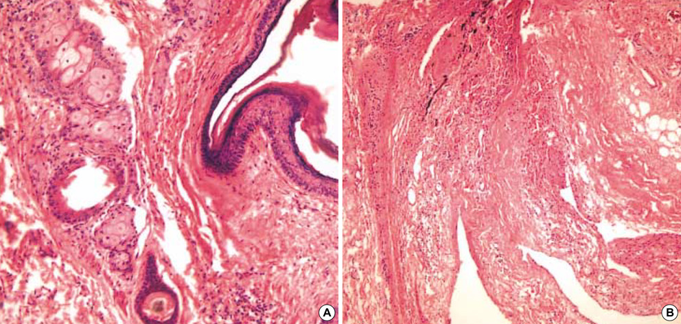
Fig. 2 Stratified squamous epithelium with skin appendage including sebaceous glands and hair follicle are noted (A, H & E, ×200). Neural tissue and adipocytes are present (B, H & E, ×100).

Fig. 3 Laboratory data after admission. DAT, direct antiglobulin test; IAT, indirect antiglobulin test.
Reference
-
1. Jung CK, Park JS, Lee EJ, Kim SH, Kwon HC, Kim JS, Roh MS, Yoon SK, Kim KH, Han JY, Kim HJ. Autoimmune hemolytic anemia in a patient with primary ovarian non-Hodgkin's lymphoma. J Korean Med Sci. 2004. 19:294–296.
Article2. Sallah S, Sigounas G, Vos P, Wan JY, Nguyen NP. Autoimmune hemolytic anemia in patients with non-Hodgkin's lymphoma: characteristics and significance. Ann Oncol. 2000. 11:1571–1577.
Article3. Glorieux I, Chabbert V, Rubie H, Baunin C, Gaspard MH, Guitard J, Duga I, Suc A, Puget C, Robert A. Autoimmune hemolytic anemia associated with a mature ovarian teratoma. Arch Pediatr. 1998. 5:41–44.4. Cobo F, Pereira A, Nomdedeu B, Gallart T, Ordi J, Torne A, Monserrat E, Rozman C. Ovarian dermoid cyst-associated autoimmune hemolytic anemia: a case report with emphasis on pathogenic mechanisms. Am J Clin Pathol. 1996. 105:567–571.5. Buonanno G, Gonnella F, Pettinato G, Castaldo C. Autoimmune hemolytic anemia and dermoid cyst of the mesentery. A case report. Cancer. 1984. 54:2533–2536.
Article6. Young RH. New and unusual aspects of ovarian germ cell tumors. Am J Surg Pathol. 1993. 17:1210–1224.
Article7. Neff AT. Greer JP, Foerster J, Lukens JN, Rodgers GM, Paraskevas F, Glader B, editors. Autoimmune hemolytic anemias. Wintrobe's Clinical Hematology. 2004. Vol 2:2nd ed. Philadelphia: Lippincott;1157–1182.8. Spira MA, Lynch EC. Autoimmune hemolytic anemia and carcinoma: An unusual association. Am J Med. 1979. 67:753–758.
Article
- Full Text Links
-
- Actions
-
Cited
- CITED
-
- Close
- Share
-
- Similar articles
-
- Two Cases of Autoimmune Hemolytic Anemia in Ulcerative Colitis
- A Case of Autoimmune Hemolytic Anemia after Fludarabine Treatment in Waldenstrom Macroglobulinemia
- Quetiapine Induced Autoimmune Hemolytic Anemia in a Child Patient: A Case Report
- A Case of Autoimmune Hemolytic Anemia Associated with Systemic Lupus Erythematosus
- Two Cases of Autoimmune Hemolytic Anemic
