Flow Cytometric Human Leukocyte Antigen-B27 Typing with Stored Samples for Batch Testing
- Affiliations
-
- 1Department of Clinical Pathology, Kyungpook National University School of Medicine, Daegu, Korea. wondi@knu.ac.kr
- KMID: 1781322
- DOI: http://doi.org/10.3343/alm.2013.33.3.174
Abstract
- BACKGROUND
Flow cytometry (FC) HLA-B27 typing is still used extensively for the diagnosis of spondyloarthropathies. If patient blood samples are stored for a prolonged duration, this testing can be performed in a batch manner, and in-house cellular controls could easily be procured. In this study, we investigated various methods of storing patient blood samples.
METHODS
We compared four storage methods: three methods of analyzing lymphocytes (whole blood stored at room temperature, frozen mononuclear cells, and frozen white blood cells [WBCs] after lysing red blood cells [RBCs]), and one method using frozen platelets (FPLT). We used three ratios associated with mean fluorescence intensities (MFI) for HLAB27 assignment: the B27 MFI ratio (sample/control) for HLA-B27 fluorescein-5-isothiocyanate (FITC); the B7 MFI ratio for HLA-B7 phycoerythrin (PE); and the ratio of these two ratios, B7/B27 ratio.
RESULTS
Comparing the B27 MFI ratios of each storage method for the HLA-B27+ samples and the B7/B27 ratios for the HLA-B7+ samples revealed that FPLT was the best of the four methods. FPLT had a sensitivity of 100% and a specificity of 99.3% for HLA-B27 assignment in DNA-typed samples (N=164) when the two criteria, namely, B27 MFI ratio >4.0 and B7/B27 ratio <1.5, were used.
CONCLUSIONS
The FPLT method was found to offer a simple, economical, and accurate method of FC HLA-B27 typing by using stored patient samples. If stored samples are used, this method has the potential to replace the standard FC typing method when used in combination with a complementary DNA-based method.
MeSH Terms
Figure
-
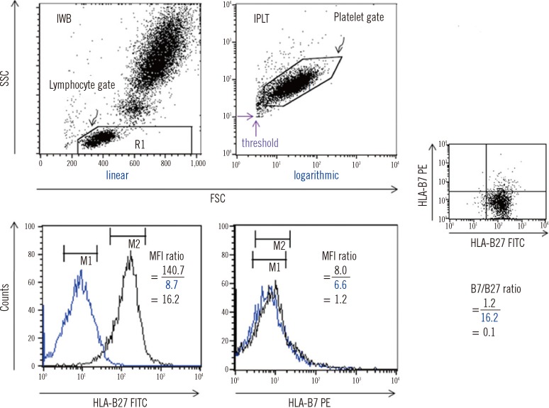
Fig. 1 FC HLA-B27 typing using the two immediate methods. The black and blue peaks denote a test sample (HLA-B27+) and a negative control, respectively. On each FSC vs. SSC plot, a lymphocyte gate was drawn for IWB using linear amplification, and a platelet gate was drawn for IPLT using logarithmic amplification. For IPLT, the FSC and SSC threshold values (the two purple arrows) were set in order to effectively collect the platelet signals by eliminating unwanted events. MFI values were obtained as the geometric means of the peaks on the HLA-B27 FITC or B7 PE histogram of the gated lymphocytes or platelets. An example of the B27 and B7 MFI ratios obtained by analyzing lymphocytes and the corresponding B7/B27 ratio is presented.Abbreviations: FC, flow cytometry; IWB, immediate whole blood; IPLT, immediate platelets; MFI, mean fluorescence intensity; FITC, fluorescein-5-isocyanate; PE, phycoerythrin; FSC, forward scatter; SSC, side scatter.
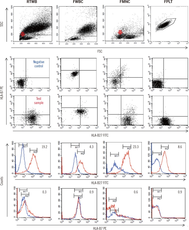
Fig. 2 Representative examples of the four storage methods. The HLA-B phenotype of the test sample (red line) was HLA-B27, B62. The negative control (blue line) for FMNC was a random donor pool showing a broad peak. MFI ratios are presented in the right upper region of each histogram. Red arrows indicate dead cells detected during or after storage by FMNC or RTWB.Abbreviations: RTWB, room temperature-stored whole blood; FWBC, frozen WBCs after lysing RBCs; FMNC, frozen mononuclear cells; FPLT, frozen platelets; FSC, forward scatter; SSC, side scatter; FITC, fluorescein-5-isocyanate; PE, phycoerythrin; MFI, mean fluorescence intensity.
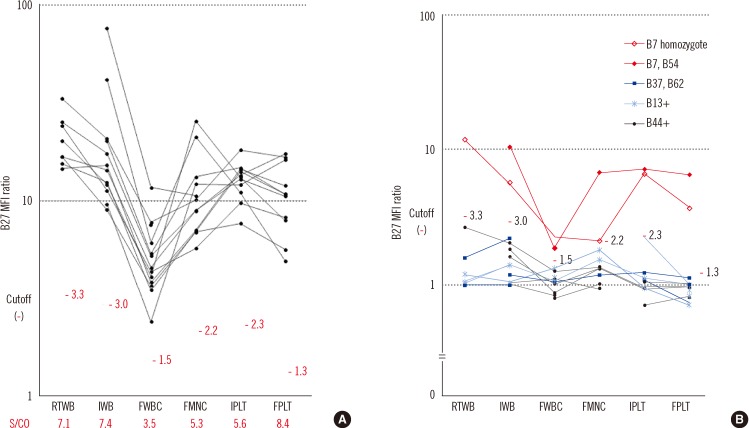
Fig. 3 Changes in B27 MFI ratios by storage method in HLA-B27+ (N=11, panel A) and B7 CREG or HLA-B44+ (N=11, panel B) samples. The cutoff B27 MFI ratio for each FC typing method was calculated in Experiment B after excluding two HLA-B7+ samples. Sample/cutoff (S/CO) ratios for each FC typing method are presented in panel A.Abbreviations: MFI, mean fluorescence intensity; IWB, immediate whole blood; IPLT, immediate platelets; RTWB, room temperature-stored whole blood; FMNC, frozen mononuclear cells; FWBC, frozen WBCs after lysing RBCs; FPLT, frozen platelets.
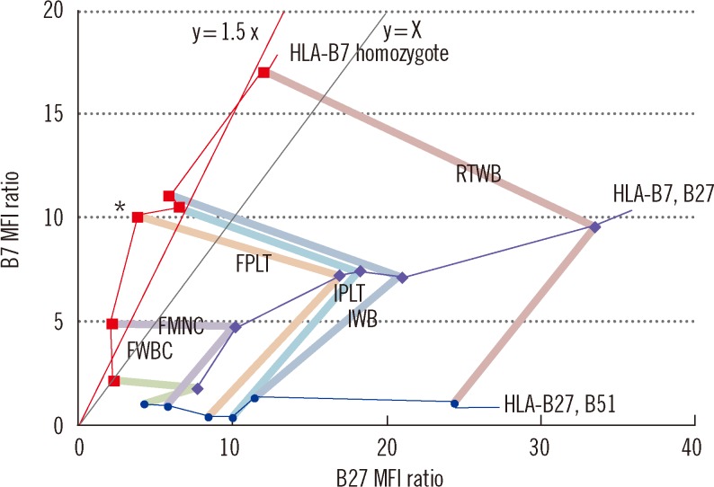
Fig. 4 Changes in B7/B27 ratios on the B27 vs. B7 MFI ratio plot according to storage method in three donors: 1) HLA-B7 homozygote (red ▪), 2) HLA-B7, B27 (purple ♦), and 3) HLA-B27, B51 (blue •). The six results obtained for a given donor are connected by thin colored lines. The three results obtained for a given FC typing method are connected by thick colored lines. The slope of the virtual straight line, which passes through the origin of the coordinate, corresponds to its B7/B27 ratio. One asterisk (*) indicates the highest B7/B27 ratio (2.7) of the six FC typing methods for a HLA-B7 homozygous donor. This value was obtained using FPLT.Abbreviationds: MFI, mean fluorescence intensity; IWB, immediate whole blood; IPLT, immediate platelets; RTWB, room temperature-stored whole blood; FMNC, frozen mononuclear cells; FWBC, frozen WBCs after lysing RBCs; FPLT, frozen platelets.
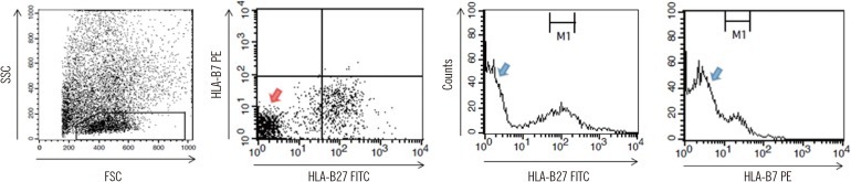
Fig. 5 An example of a caveat of using RTWB. An HLA-B27+ sample, which was held for seven days at room temperature, showed many particles with weak fluorescence (red arrow) on the FL1 vs. FL2 plot of gated lymphocytes. These particles constituted a main peak (2 blue arrows) on each fluorescence histogram, which could have been mistakenly interpreted as a negative reaction.Abbreviations: SSC, side scatter; FSC, forward scatter; PE, phycoerythrin; FITC, fluorescein-5-isocynate; M1, marker 1.
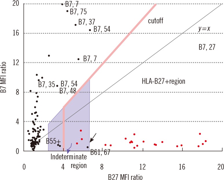
Fig. 6 B27 vs. B7 MFI ratio plot for FC HLA-B27 typing by FPLT (N=164). The results of DNA typing are indicated as red (HLA-B27+) or black (HLA-B27-) dots, or are written alongside the dots. The cutoff line (red) is composed of two straight lines: 1) x =4; and 2) y=1.5x. Dots under this broken border were assigned HLA-B27+ by FPLT. FPLT was concordant with DNA typing in all cases, except for a single false-positive case indicated by the black arrow (phenotype HLA-B61, B67). Cases in the indeterminate region (purple, shaded area) required confirmation by DNA typing for a robust determination to be reached. Refer to the DISCUSSION for a definition of the indeterminate region. There were two HLA-B27- samples in the indeterminate region, but not in the HLA-B27+ region, whose phenotypes were determined as HLA-B55+ by DNA typing.Abbreviations: MFI, mean fluorescence intensity; FPLT, frozen platelets.
Reference
-
1. Reveille JD. Major histocompatibility genes and ankylosing spondylitis. Best Pract Res Clin Rheumatol. 2006; 20:601–609. PMID: 16777585.
Article2. Seipp MT, Erali M, Wies RL, Wittwer C. HLA-B27 typing: evaluation of an allele-specific PCR melting assay and two flow cytometric antigen assays. Cytometry B Clin Cytom. 2005; 63:10–15. PMID: 15624199.
Article3. College of American Pathologists. Surveys 2011 B27-A HLA-B27 typing participant summary. 2011. Northfield: College of American Pathologists;p. 1–5. (http://www.cap.org).4. Beckman Coulter. IOTest HLA-B27-FITC/HLA-B7-PE package insert (A07739). Last visit on Jan 2013. http://www.beckmancoulter.com/wsrportal/page/itemDetails?itemNumber=A07739#2/10//0/25/1/0/asc/2/A07739///0/1//0/.5. Levering WH, Wind H, Granger V, Sintnicolaas K, Hooijkaas H, Reilly JT, et al. Long-term stabilized blood samples as controls for flow cytometric HLA-B27 screening: a feasibility study. Cytometry B Clin Cytom. 2008; 74:169–181. PMID: 18200592.
Article6. European Federation for Immunogenetics. Standards for Histocompatibility Testing. 2005. 4. Version 5.4.7. American Society for Histocompatibility and Immunogenetics. Revised Standards for Histocompatibility Testing. 2005. 12.8. Levering WH, Sintnicolaas K, Wind H, Hooijkaas H, Gratama JW. Flow cytometric screening for the HLA-B27 antigen on peripheral blood lymphocytes. Curr Protoc Cytom. 2005; 8. Chapter 6: Unit6.22.9. Coates E, Darke C. Routine HLA-B27 typing by flow cytometry: differentiation of the products of HLA-B*2702, B*2705 and B*2708. Eur J Immunogenet. 1998; 25:29–37. PMID: 9587742.
Article10. Coates E, Rees TJ, Darke C. An evaluation of Com-B27, a fluorescein-conjugated mouse anti-HLA-B27 reagent. Eur J Immunogenet. 2003; 30:271–274. PMID: 12919288.
Article11. Rodey GE, Revels K, Fuller TC. Epitope specificity of HLA class I alloantibodies: II. Stability of cross-reactive group antibody patterns over extended time periods. Transplantation. 1997; 63:885–893. PMID: 9089230.12. McFarland JG. Roback JD, Combs MR, Grossman BJ, Hillyer CD, editors. Platelet and granulocyte antigens. Technical manual. 2008. 16th ed. Bethesda: AABB;p. 525–546.13. Helmerhorst FM, ten Berge ML, van der Plas-van Dalen CM, Engelfriet CP, von dem Borne AE. Platelet freezing for serological purposes with and without a cryopreservative. Vox Sang. 1984; 46:318–322. PMID: 6375132.
Article14. National Institute for Biological Standards and Control. Platelet immunofluorescence test protocol. Last visit on Oct 2012. http://www.nibsc.ac.uk/science/diagnostics/transfusion__transplantation/platelets/resources/pift_protocol.aspx.15. Moroff G, Seetharaman S, Kurtz JW, Greco NJ, Mullen MD, Lane TA, et al. Retention of cellular properties of PBPCs following liquid storage and cryopreservation. Transfusion. 2004; 44:245–252. PMID: 14962316.
Article16. Maecker HT, Moon J, Bhatia S, Ghanekar SA, Maino VC, Payne JK, et al. Impact of cryopreservation on tetramer, cytokine flow cytometry, and ELISPOT. BMC Immunol. 2005; 6:17. PMID: 16026627.
Article17. Cho EH, Lee SG, Seok JH, Park BY, Lee EH. Evaluation of two commercial HLA-B27 real-time PCR kits. Korean J Lab Med. 2009; 29:589–593. PMID: 20046093.
Article18. McKenna RM, Takemoto SK. Improving HLA matching for kidney transplantation by use of CREGs. Lancet. 2000; 355:1842–1843. PMID: 10866435.
Article19. Porretti L, Marangoni F, Rebulla P, Sirchia G. Frozen platelet plates for platelet antibody detection and cross-match. Vox Sang. 1994; 67:52–57. PMID: 7975453.
Article20. Darke C, Coates E. One-tube HLA-B27/B2708 typing by flow cytometry using two "Anti-HLA-B27" monoclonal antibody reagents. Cytometry B Clin Cytom. 2010; 78:21–30. PMID: 19693889.
Article21. Lee SH, Choi IA, Lee YA, Park EK, Kim YH, Kim KS, et al. Human leukocyte antigen-B*2705 is the predominant subtype in the Korean population with ankylosing spondylitis, unlike in other Asians. Rheumatol Int. 2008; 29:43–46. PMID: 18493767.
Article
- Full Text Links
-
- Actions
-
Cited
- CITED
-
- Close
- Share
-
- Similar articles
-
- Comparison of HLA-B27 typing methods -PCR-SSP, microlymphocytotoxicity, and flow cytometry
- Evaluation of a Korean HLA-B27 typing tray
- Comparison of the Flowcytometric HLA-B27 Determination Methods
- Annual Report of Korean Association of External Quality Assessment Service on Histocompatibility Testing (2018)
- The Comparison of Duration to Maintain Cell Viability for HLA-B27 Test According to Anticoagulants
