Ocular Sarcoidosis in a Korean Population
- Affiliations
-
- 1Department of Ophthalmology, Asan Medical Center, College of Medicine, University of Ulsan, Seoul, Korea. yhyoon@amc.seoul.kr
- 2Department of Pulmonary Medicine, Asan Medical Center, College of Medicine, University of Ulsan, Seoul, Korea.
- 3Department of Ophthalmology and Neuroscience, Johns Hopkins University School of Medicine, Baltimore, Maryland, U.S.A.
- KMID: 1779156
- DOI: http://doi.org/10.3346/jkms.2009.24.3.413
Abstract
- The aim of current study was to evaluate the incidence and characteristics of ocular sarcoidosis in a Korean population. We conducted a retrospective study of 104 consecutive patients with biopsy-proven sarcoidosis seen at Asan Medical Center in Seoul, Korea, from 1993 to 2007. Medical records, photographs, and fluorescein angiograms were reviewed. Of 104 patients, 22 (21%) had intraocular involvement with female predominance (86%, M:F=3:19). Of the 39 eyes with ocular involvement, 16 (41%) eyes had isolated anterior uveitis, 12 (31%) eyes had intermediate uveitis, 6 eyes (15%) had panuveitis with retinal vasculitis, and 5 (13%) eyes had panuveitis with punched multifocal choroiditis. Mean duration of ophthalmologic follow-up was 62 months. All ocular inflammation was well managed with topical steroid and/or systemic steroid with relatively good final visual outcomes. Ocular complications such as cataract (12 eyes, 30%), glaucoma (6 eyes, 15%), vitreous opacity (1 eye, 3%), cystoid macular edema (3 eyes, 7%), neovascularization (2 eye, 5%), and epiretinal membrane (4 eye, 10%) were related to ocular sarcoidosis. In Korea, where sarcoidosis is very rare, our study indicates relatively low ocular and predominantly non posterior segment involvement with relatively good visual prognosis.
Keyword
MeSH Terms
-
Adult
Age Factors
Asian Continental Ancestry Group
Choroiditis/diagnosis
Eye Diseases/*diagnosis/epidemiology/therapy
Female
Fluorescein Angiography
Humans
Male
Middle Aged
Retinal Vasculitis/diagnosis
Retrospective Studies
Sarcoidosis/*diagnosis/epidemiology/therapy
Steroids/therapeutic use
Uveitis, Anterior/diagnosis
Uveitis, Intermediate/diagnosis
Figure
-
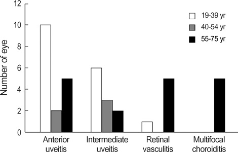
Fig. 1 Comparision of age among four ordered types of uveitis. While anterior or intermediate uveitis tend to occur in younger age group, all 3 patients (5 eyes) showing 'panuveitis with punched multifocal choroiditis' were over 55 yr old.
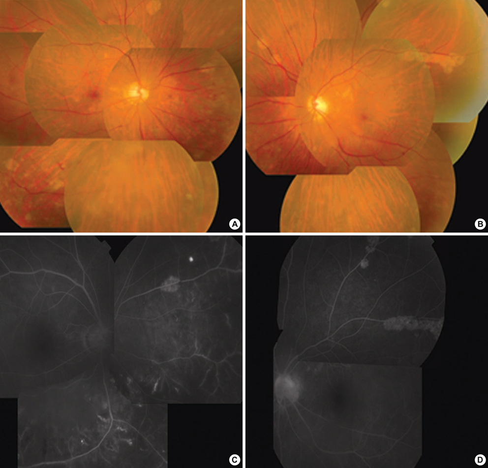
Fig. 2 Patient No. 17. Fundus photographs (A, B) show retinal vasculitis and multiple depigmented peripheral lesions. Fluorescence angiography (FAG) (C, D) shows late fluorescein leakage from retinal vessels with adjacent capillary nonperfusion.
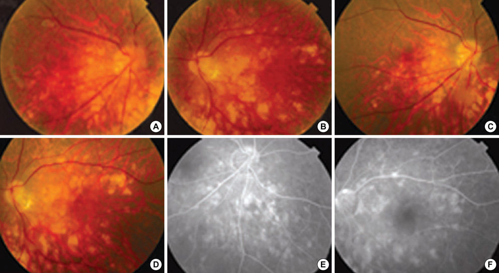
Fig. 3 Patient No. 21. Fundus photographs show subacute multifocal choroiditis with accompanying vitreous opacity at initial presentation (A, B) and residual choroiditis accompanying atrophic change at one month later (C, D). Fluorescence angiography of right inferonasal aspect and left posterior pole of retina at one month later from initial presentation (E, F) show atrophic choroidal changes.
Cited by 2 articles
-
Peripapillary Granuloma with Optic Nerve Head Involvement Associated with Sarcoidosis
In Kwon Chung, Jonghyun Lee, Joo Youn Shin
Korean J Ophthalmol. 2019;33(4):389-391. doi: 10.3341/kjo.2018.0112.Ocular Manifestations of Sarcoidosis: An Ophthalmologist's View
Yun Taek Kim
Hanyang Med Rev. 2016;36(3):168-173. doi: 10.7599/hmr.2016.36.3.168.
Reference
-
1. Hunter DG, Foster CS. Albert DM, Jakobiec FA, editors. Ocular manifestations of sarcoidosis. Principles and Practice of Opthalmology. 1994. Philadelphia: WB Saunders;1217–1224.2. Obenauf CD, Shaw HE, Sydnor CF, Klintworth GK. Sarcoidosis and its ophthalmic manifestations. Am J Ophthalmol. 1978. 86:648–655.
Article3. James DG, Anderson R, Langley D, Ainslie D. Ocular sarcoidosis. Br J Ophthalmol. 1964. 48:461–470.
Article4. Karma A. Ophthalmic changes in sarcoidosis. Acta Ophthalmol Suppl. 1979. 141:1–94.5. Jabs DA, Johns CJ. Ocular involvement in chronic sarcoidosis. Am J Ophthalmol. 1986. 102:297–301.
Article6. Rybicki BA, Major M, Popovich J Jr, Maliarik MJ, Iannuzzi MC. Racial differences in sarcoidosis incidence: a 5-year study in a health maintenance organization. Am J Epidemiol. 1997. 145:234–241.
Article7. Hunninghake GW, Costabel U, Ando M, Baughman R, Cordier JF, du Bois R, Eklund A, Kitaichi M, Lynch J, Rizzato G, Rose C, Selroos O, Semenzato G, Sharma OP. ATS/ERS/WASOG statement on sarcoidosis. American Thoracic Society/European Respiratory Society/ World Association of Sarcoidosis and other Granulomatous Disorders. Sarcoidosis Vasc Diffuse Lung Dis. 1999. 16:149–173.8. Kim DS. Sarcoidosis in Korea: report of the second nationwide survey. Sarcoidosis Vasc Diffuse Lung Dis. 2001. 18:176–180.9. Hosoda Y, Yamaguchi M, Hiraga Y. Global epidemiology of sarcoidosis. What story do prevalence and incidence tell us? Clin Chest Med. 1997. 18:681–694.10. BenEzra D, Rorrester JV, Nussenblat RB. Uveitis Scoring System. Berlin: Springer-Verlag;1–13.11. James DG, Neville E, Langley DA. Ocular sarcoidosis. Trans Ophthalmol Soc U K. 1976. 96:133–139.
Article12. Rothova A, Alberts C, Glasius E, Kijlstra A, Buitenhuis HJ, Breebaart AC. Risk factors for ocular sarcoidosis. Doc Ophthalmol. 1989. 72:287–296.
Article13. Iwata K, Nanba K, Sobue K, Abe H. Ocular sarcoidosis: evaluation of intraocular findings. Ann N Y Acad Sci. 1976. 278:445–454.
Article14. Yamaguchi M, Hosoda Y, Sasaki R, Aoki K. Epidemiological study on sarcoidosis in Japan. Recent trends in incidence and prevalence rates and changes in epidemiological features. Sarcoidosis. 1989. 6:138–146.15. Ohara K, Okubo A, Sasaki H, Kamata K. Intraocular manifestations of systemic sarcoidosis. Jpn J Ophthalmol. 1992. 36:452–457.16. Lobo A, Barton K, Minassian D, du Bois RM, Lightman S. Visual loss in sarcoid-related uveitis. Clin Experiment Ophthalmol. 2003. 31:310–316.
Article17. Khalatbari D, Stinnett S, McCallum RM, Jaffe GJ. Demographic-related variations in posterior segment ocular sarcoidosis. Ophthalmology. 2004. 111:357–362.
Article18. Thorne JE, Brucker AJ. Choroidal white lesions as an early manifestation of sarcoidosis. Retina. 2000. 20:8–15.
Article19. Lardenoye CW, Van der Lelij A, de Loos WS, Treffers WF, Rothova A. Peripheral multifocal chorioretinitis: a distinct clinical entity? Ophthalmology. 1997. 104:1820–1826.20. Hershey JM, Pulido JS, Folberg R, Folk JC, Massicotte SJ. Non-caseating conjunctival granulomas in patients with multifocal choroiditis and panuveitis. Ophthalmology. 1994. 101:596–601.
Article21. Dana MR, Merayo-Lloves J, Schaumberg DA, Foster CS. Prognosticators for visual outcome in sarcoid uveitis. Ophthalmology. 1996. 103:1846–1853.
Article22. Bardelli AM, Barberi L, Vanni M, Mazzera L, Traversi C. Eye involvement in sarcoidosis: survey of 197 patients. Sarcoidosis. 1993. 10:158–159.23. Sugisaki K, Yamaguchi T, Nagai S, Ohmiti M, Takenaka S, Morimoto S, Ishihara M, Tachibana T, Tsuda T. Clinical characteristics of 195 Japanese sarcoidosis patients treated with oral corticosteroids. Sarcoidosis Vasc Diffuse Lung Dis. 2003. 20:222–226.
