J Korean Med Sci.
2005 Jun;20(3):427-432. 10.3346/jkms.2005.20.3.427.
An Experimental Animal Model of Fulminant Hepatic Failure in Pigs
- Affiliations
-
- 1Department of Surgery, Seoul National University College of Medicine, Seoul, Korea. kulee@plaza.snu.ac.kr
- 2Department of Surgery, The 2nd Affiliated Hospital of Harbin Medical University, Harbin, PR China.
- KMID: 1778501
- DOI: http://doi.org/10.3346/jkms.2005.20.3.427
Abstract
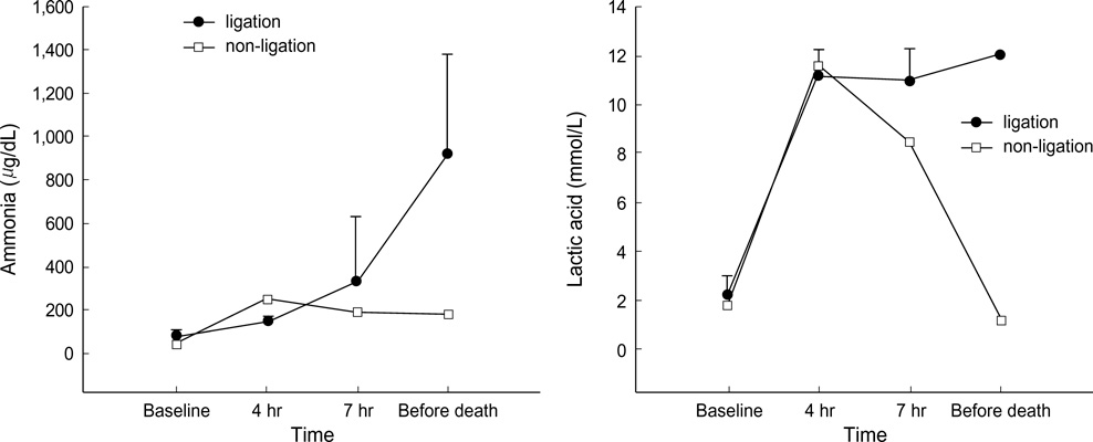
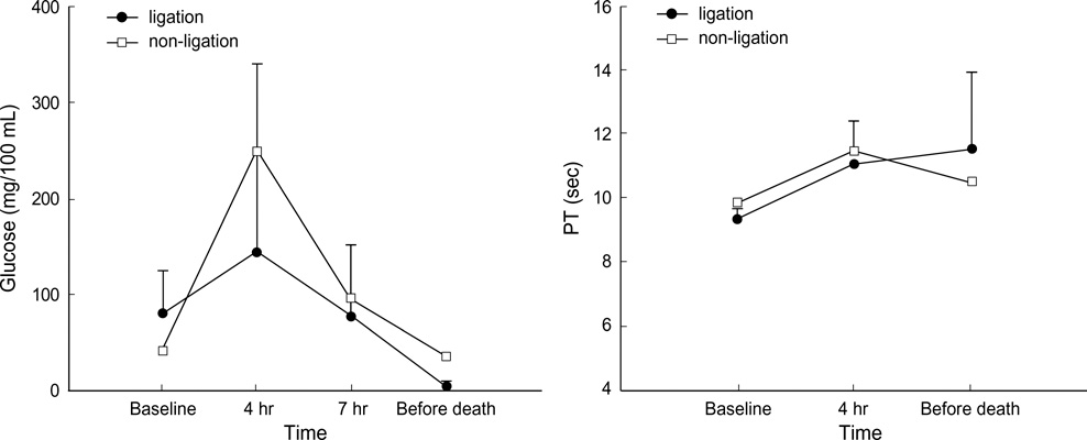
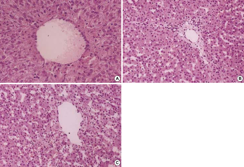
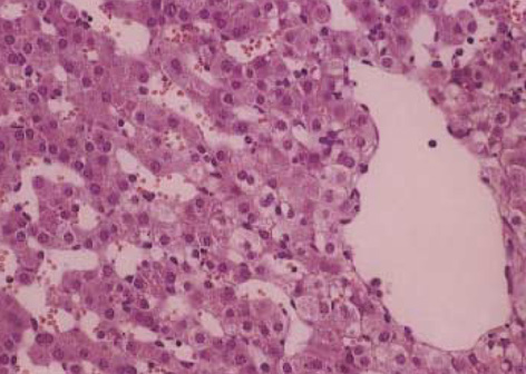
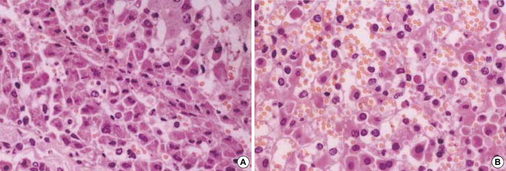
- The objective of this study was to develop an experimental animal model of fulminant hepatic failure to test the efficacy of the bioartificial liver system. The portal vein and the hepatic artery were clamped intermittently and then the hepatic artery was ligated (ligation group, n=5). Pigs whose hepatic arteries were not ligated after clamping were assigned to the non-ligation group (n=5). The biochemical changes in blood, histologic alterations of the liver and neurologic examination for pigs were checked up. All animals died within 17 hr in the ligation group. On the other hand, all animals survived more than 7 days in the non-ligation group. In the ligation group, the levels of ammonia, lactic acid and creatinine showed a progressively increasing pattern. Prothrombin time was also prolonged gradually. Cytoplasmic condensation and nuclear pyknosis of hepatocytes were detected histologically at autopsy. Neurologic findings such as decreased pain sensation, tachypnea and no light reflex of pupils were observed. The findings shown in the ligation group are similar to the clinical features of fulminant hepatic failure in human and this animal model is reproducible. Therefore, this can be a suitable animal model to evaluate the efficacy of the bioartificial liver system for treating fulminant hepatic failure.
MeSH Terms
-
Acidosis/etiology/prevention & control
Ammonia/blood
Animals
Aspartate Aminotransferases/blood
Bilirubin/blood
Blood Glucose/metabolism
Blood Urea Nitrogen
Comparative Study
Creatinine/blood
*Disease Models, Animal
Female
Hepatic Artery/surgery
Lactic Acid/blood
Ligation/adverse effects
Liver Failure, Acute/blood/*pathology
Portal Vein/surgery
Potassium/blood
Prothrombin Time
Research Support, Non-U.S. Gov't
Sodium Bicarbonate/pharmacology
Swine
Figure
Reference
-
1. Lee WM. Acute liver failure. Am J Med. 1994. 96:3S–9S.
Article2. Adam R, Cailliez V, Majno P, Karam V, McMaster P, Caine RY, O'Grady J, Pichlmayr R, Neuhaus P, Otte JB, Hoeckerstedt K, Bismuth H. Normalised intrinsic mortality risk in liver transplantation: European Liver Transplant Registry study. Lancet. 2000. 356:621–627.
Article3. Lee WM. Acute liver failure. N Engl J Med. 1993. 329:1862–1872.
Article4. Terblanche J, Hickman R. Animal models of fulminant hepatic failure. Dig Dis Sci. 1991. 36:770–774.
Article5. Benoist S, Sarkis R, Baudrimont M, Delelo R, Robert A, Vaubourdolle M, Balladur P, Calmus Y, Capeau J, Nordlinger B. A reversible model of acute hepatic failure by temporary hepatic ischemia in the pig. J Surg Res. 2000. 88:63–69.
Article6. de Groot GH, Reuvers CB, Schalm SW, Boks AL, Terpstra OT, Jeekel H, ten Kate FW, Bruinvels J. A reproducible model of acute hepatic failure by transient ischemia in the pig. J Surg Res. 1987. 42:92–100.
Article7. Filipponi F, Fabbri LP, Marsili M, Falcini F, Benassai C, Nucera M, Romagnoli P. A new surgical model of acute liver failure in the pig: experimental procedure and analysis of liver injury. Eur Surg Res. 1991. 23:58–64.
Article8. Sielaff TD, Hu MY, Rollins MD, Bloomer JR, Amiot B, Hu WS, Cerra FB. An anesthetized model of lethal canine galactosamine fulminant hepatic failure. Hepatology. 1995. 21:796–804.
Article9. Kalpana K, Ong HS, Soo KC, Tan SY, Prema Raj J. An improved model of galactosamine-induced fulminant hepatic failure in the pig. J Surg Res. 1999. 82:121–130.
Article10. Diaz-Buxo JA, Blumenthal S, Hayes D, Gores P, Gordon B. Galactosamine-induced fulminant hepatic necrosis in unanesthetized canines. Hepatology. 1997. 25:950–957.
Article11. Kelly JH, Koussayer T, He DE, Chong MG, Shang TA, Whisennand HH, Sussman NL. An improved model of acetaminophen-induced fulminant hepatic failure in dogs. Hepatology. 1992. 15:329–335.
Article12. Mourelle M, Villlalon C, Amezcua JL. Protective effect of colchicine on acute liver damage induced by carbon tetrachloride. J Hepatol. 1988. 6:337–342.
Article13. Starzl TE, Demetris AJ. Liver transplantation: a 31-year perspective. Part I. Curr Probl Surg. 1990. 27:49–116.14. Nordlinger B, Douvin D, Javaudin L, Bloch P, Aranda A, Boschat M, Huguet C. An experimental study of survival after two hours of normothermic hepatic ischemia. Surg Gynecol Obstet. 1980. 150:859–864.15. Harris KA, Wallace AC, Wall WJ. Tolerance of the liver to ischemia in the pig. J Surg Res. 1982. 33:524–530.
Article16. Nitta N, Yamamoto S, Ozaki N, Morimoto T, Mori K, Yamaoka Y, Ozawa K. Is the deterioration of liver viability due to hepatic warm ischemia or reinflow of pooled-portal blood in intermittent portal triad cross-clamping? Res Exp Med (Berl). 1988. 188:341–350.
Article17. Li YM, Qin ZY, Kang YA, Ji ZZ, Yang WB, Xiang GA, Fang ZW. Injurious effects of hepatic arterial ischemia on hepatic and biliary ductal tissues in canine liver autografts. Chin J Organ Transplant. 1996. 17:28–30.18. Ferenci P. Brain dysfunction in fulminant hepatic failure. J Hepatol. 1994. 21:487–490.
Article19. Hillenbrand P, Parbhoo SP, Jedrychowski A, Sherlock S. Significance of intravascular coagulation and fibrinolysis in acute hepatic failure. Gut. 1974. 15:83–88.
Article
- Full Text Links
- Actions
-
Cited
- CITED
-
- Close
- Share
- Similar articles
-
- Development of Fulminant Hepatic Failure in Pigs
- Three Cases of Fulminant Hepatic Failure due to Congestive Heart Failure
- Animal Models of Acute Hepatic Failure
- A case of fulminant hepatic failure complicating hepatitis A virus superinfection in a hepatitis B virus carrier
- A Case of Fulminant Hepatic Failure Secondary to Hepatic Metastasis of Small Cell Lung Carcinoma

