Marginal Zone B-cell Lymphoma of MALT in Small Intestine Associated with Amyloidosis: A Rare Association
- Affiliations
-
- 1Department of Pathology, Gil Medical Center, Gachon University, School of Medicine and Science, Incheon, Korea. hicho@gilhospital.com
- KMID: 1777871
- DOI: http://doi.org/10.3346/jkms.2011.26.5.686
Abstract
- A 62-yr-old man presented with a 5-yr history of intermittent abdominal distention and pain. These symptoms persisted for several months and subsided without treatment. A diagnosis of suspected small bowel lymphoma was made based on plain radiograph and computerized tomogram findings, and he was referred to our institution for further evaluation. Segmental resection of the small intestine was performed and the diagnosis of marginal zone B-cell lymphoma associated with amyloidosis was made. This is the first case of marginal zone B-cell lymphoma of mucosa-associated lymphoid tissue (MALT) in the small intestine associated with amyloidosis in Korea.
MeSH Terms
Figure
-

Fig. 1 Histologic features of a resection specimen of small intestine. (A) The intestinal wall was thickened by a double process. Here, amorphous acidophilic material in the submucosa and intestinal wall were mixed with lymphoid infiltrate (H&E, scanning view). (B) High power view (H&E, × 40) and apple-green bi-refringence under polarized light is seen Congo-red staining of amyloid (inset). (C) Diffuse proliferative lymphoid infiltrate through the intestinal wall up to the serosa (H&E, scanning view). (D) The lymphoid infiltrate was composed mainly of small lymphoid cells (H&E, × 400).
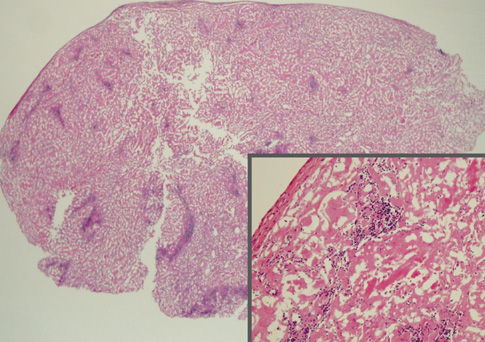
Fig. 2 Histologic features of mesenteric lymph node biopsy. Amyloid deposits are dense admixed with some aggregates of lymphoid cells. Islands of lymphoid infiltration are composed of small lymphoid cells, some of which show plasmacytic differentiation (H&E, scanning view and × 100-inset).

Fig. 3 The immunohistochemical findings. The lymphoid infiltrate was positive for CD20, (A) Ki-67 labeling index was low (less than 5%), (B) kappa light chain-producing plasma cells were detected, (C) with the exclusion of lambda light chain-producing plasma cells, (D) (immunoperoxidase, × 400).
Reference
-
1. Briggs GW. Amyloidosis. Ann Intern Med. 1961. 55:943–957.2. Legge DA, Carlson HC, Wollaeger EE. Roentgenologic appearance of systemic amyloidosis involving gastrointestinal tract. Am J Roentgenol Radium Ther Nucl Med. 1970. 110:406–412.3. Baldewijns M, Ectors N, Verbeeck G, Janssens J, De Schepper J, Ponette E, Geboes K, Desmet V. Intermittent subobstruction and cholestasis as complications of duodenal amyloid tumours. Gastroenterol Clin Biol. 1995. 19:218–221.4. Hamaya K, Kitamura M, Doi K. Primary amyloid tumors of the jejunum producing intestinal obstruction. Acta Pathol Jpn. 1989. 39:207–211.5. Hauben E, Fierens H, Heylen H, Van Marck E. Localized amyloid tumour of the duodenum: a case report. Acta Gastroenterol Belg. 1997. 60:304–305.6. Peny MO, Debongnie JC, Haot J, Van Gossum A. Localized amyloid tumor in small bowel. Dig Dis Sci. 2000. 45:1850–1853.7. Arista Nasr J, Lome-Maldonado C. Diffuse small lymphoplasmacytic lymphoma of the GI tract associated with massive intestinal amyloidosis. Rev Invest Clin. 1993. 45:71–75.8. Caulet S, Robert I, Bardaxoglou E, Noret P, Tas P, Le Prise Y, Launois B, Ramee MP. Malignant lymphoma of mucosa associated lymphoid tissue: a new etiology of amyloidosis. Pathol Res Pract. 1995. 191:1203–1207.9. Das K, Ghoshal UC, Jain M, Rastogi A, Tiwari S, Pandey R. Primary gastric lymphoma and Helicobacter pylori infection with gastric amyloidosis. Indian J Gastroenterol. 2005. 24:220–222.10. Goteri G, Ranaldi R, Pileri SA, Bearzi I. Localized amyloidosis and gastrointestinal lymphoma: a rare association. Histopathology. 1998. 32:348–355.11. Ranaldi R, Goteri G, Santinelli A, Rezai B, Pileri S, Poggi S, Bearzi I. Centrocytic-like lymphoma associated with localized amyloidosis of the large intestine. Virchows Arch. 1994. 425:327–330.12. Wright JR, Calkins E, Humphrey RL. Potassium permanganate reaction in amyloidosis. A histologic method to assist in differentiating forms of this disease. Lab Invest. 1977. 36:274–281.13. Kim SY, Bang BK, Park CW, Kim KW, Yun SR, Han CM, Park YH, Ahn SJ, Park SY, Kim HJ, Suh KS, Park KK. An unusual case of AA type amyloidosis in lymphoma. Korean J Nephrol. 1999. 18:808–814.
- Full Text Links
-
- Actions
-
Cited
- CITED
-
- Close
- Share
-
- Similar articles
-
- A case with mucosa-associated lymphoid tissue(MALT) lymphoma and tuberculous enteritis at jejunum
- A Case of Primary Cutaneous Marginal Zone B-cell Lymphoma
- A Case of Primary Pulmonary Extranodal Marginal Zone B-Cell Lymphoma of the MALT Type
- A Case of Mucosa-Associated Lymphoid Tissue Lymphoma in Nasopharynx and Thyroid Gland
- Gastrointestinal Lymphoma
