Screen-Film Mammography and Soft-Copy Full-Field Digital Mammography: Comparison in the Patients with Microcalcifications
- Affiliations
-
- 1Department of Radiology and the Center for Imaging Science, Samsung Medical Center, Sungkyunkwan University School of Medicine, Korea. bkhan@smc.samsung.co.kr
- 2Department of Surgery, Samsung Medical Center, Sungkyunkwan University School of Medicine, Korea.
- KMID: 1777278
- DOI: http://doi.org/10.3348/kjr.2005.6.4.214
Abstract
OBJECTIVE
We wanted to compare the ability of screen-film mammography (SFM) and soft-copy full-field digital mammography (s-FFDM) on two different monitors to detect and characterize microcalcifications. MATERIALS AND METHODS: The images of 40 patients with microcalcifications (three patients had malignant lesion and 37 patients had benign lesion), who underwent both SFM and FFDM at an interval of less than six months, were independently evaluated by three readers. Three reading sessions were undertaken for SFM and for FFDM on a mammography-dedicated review workstation (RWS, 2K x 2.5K), and for FFDM on a high-resolution PACS monitor (1.7K x 2.3K). The image quality, breast composition and the number and conspicuity of the microcalcifications were evaluated using a three-point rating method, and the mammographic assessment was classified into 4 categories (normal, benign, low concern and moderate to great concern). RESULTS: The image quality, the number and conspicuity of the microcalcifications by s-FFDM (on the RWS, PACS and both) were superior to those by SFM in 85.0%, 80.0% and 52.5% of the cases, respectively (p < 0.01), and those by the s-FFDM on the two different monitors were similar in 15.0%, 12.5% and 35.0% of the cases, respectively (p > 0.01). The mammographic assessment category for the microcalcifications in the three reading sessions was similar. CONCLUSION: s-FFDM gives a superior image quality to SFM and it is better at evaluating microcalcifications. In addition, s-FFDM with the PACS monitor is comparable to s-FFDM with the RWS for evaluating microcalcifications.
Keyword
MeSH Terms
Figure
-
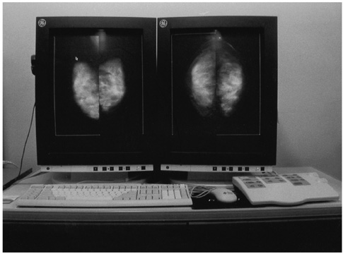
Fig. 1 Soft-copy workstation for digital mammography. The figure shows the soft-copy display system using a 2×2.5 K mammography-dedicated review workstation (General Electric Medical Systems, Buc, France).
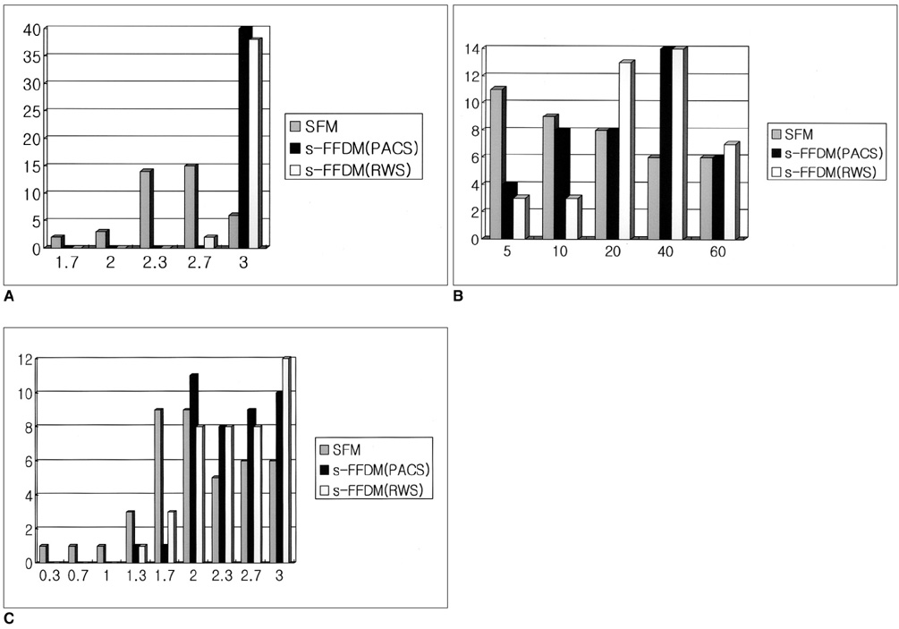
Fig. 2 Distribution of the results of the comparison between the full-field digital mammography on a PACS monitor and the full-field digital mammography on a review workstation by three readers. Image quality (A), number (B) and conspicuity (C) of microcalcifications in screen-film mammography versus full-field digital mammography.
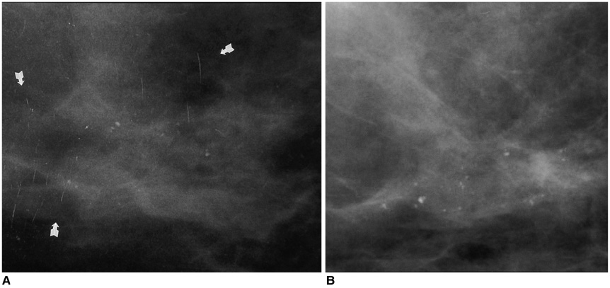
Fig. 3 Comparison of image quality. Scratch and dust artifacts are noted in A and it is difficult to differentiate the microcalcification from the dust in A, resulting in superior image quality of the full-field digital mammography (B).
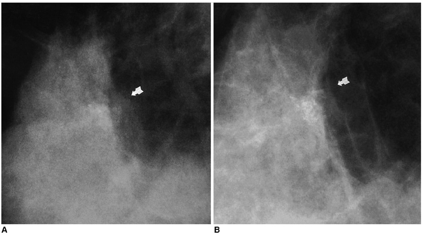
Fig. 4 Comparison of the number and conspicuity of the microcalcifications. Many more calcifications with a clear margin are noted in the full-field digital mammography (B) than in a screen-film mammography (A).
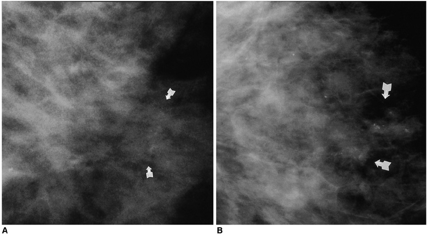
Fig. 5 A 48-year-old woman with an increased number of clustered microcalcifications in the left breast. She underwent breast-conserving surgery for an infiltrating ductal carcinoma of the right breast two years ago. The ACR BI-RADS mammographic assessment category of the microcalcification was more consistent with the pathologic result in the full-field digital mammography (B) than in the screen-film mammography (A), as assessed by all three interpreters. These microcalcifications were confirmed to be mucocele-like tumor with atypical ductal hyperplasia by a stereotaxic mammotome biopsy. A and B were performed at the same time.
Reference
-
1. Sickels EA. Mammographic features of 300 consecutive nonpalpable breast cancers. AJR Am J Roentgenol. 1986. 46:661–663.2. Feig SA, Yaffe MJ. Digital mammography, computer-aided diagnosis, and telemammography. Radiol Clin North Am. 1995. 33:1205–1230.3. Obenauer S, Luftner-Nagel S, von Heyden D, Munzel U, Baum F, Grabbe E. Screen film vs. full-field digital mammography: image quality, detectability and characterization of lesions. Eur Radiol. 2002. 12:1697–1702.4. Hermann KP, Hundertmark C, Funke M, Brenndorff AV, Grabbe E. Digital mammography in direct magnification technique using a large-area amorphous silicon X-ray detector. Rofo. 1999. 170:503–506.5. Pisano ED, Cole EB, Kistner EO, Muller KE, Hemminger BM, Brown ML, et al. Interpretation of digital mammograms: comparison of speed and accuracy of soft-copy versus printed-film display. Radiology. 2002. 223:483–488.6. Fischer U, Baum F, Obenauer S, Luftner-Nagel S, Heyden D, Vosshenrich R, et al. Comparative study in patients with microcalcifications: full-field digital mammography vs. screen-film mammography. Eur Radiol. 2002. 12:2679–2683.7. American College of Radiology. Breast imaging reporting and data system (BI-RADSTM). 1998. 3rd ed. Reston, Va.: American College of Radiology.8. Brown H, Prescott R. Applied mixed model in medicine. 1999. New York: John Wiley & Sons;199–260.9. Shtern F. Digital mammography and related technologies: a perspective from the national cancer institute. Radiology. 1992. 183:629–630.10. Kheddache S, Thilander-Klang A, Lanhede B, Mansson LG, Bjurstam N, Ackerholm P, et al. Storage phosphor and film-screen mammography: performance with different mammographic techniques. Eur Radiol. 1999. 9:591–597.11. Funke M, Hermann KP, Breiter N, Hundertmark C, Sachs J, Gruhl T, et al. Digital storage phosphor mammography in a magnification technic: experimental studies for spatial resolution and for detection of microcalcifications. Rofo. 1997. 167:174–179.12. Lewin JM, Hendrick RE, Dorsi CJ, Isaacs PK, Moss LJ, Karellas A, et al. Comparison of full-field digital mammography with screen-film mammography for cancer detection: results of 4945 paired examinations. Radiology. 2001. 218:873–880.13. Venta LA, Hendrick RE, Adler YT, DeLeon P, Mengoni PM, Scharl AM, et al. Rates and causes of disagreement in interpretation of full-field digital mammography and film-screen mammography in a diagnostic setting. AJR Am J Roentgenol. 2000. 176:1241–1248.14. Hermann KP, Obenauer S, Grabbe E. Radiation exposure in full-field digital mammography with a flat-panel X-ray detector based on amorphous silicon in comparison with conventional screen-film mammography. Rofo. 2000. 172:940–945.15. Leung JW. New modalities in breast imaging: digital mammography, positron emission tomography, and sestamibi scintimammography. Radiol Clin North Am. 2002. 40:467–482.16. James JJ. The current status of digital mammography. Clin Radiol. 2004. 59:1–10.17. Hayt DB, Alexander S, Drakakis J, Berdebes N. Filmless in 60 days: the impact of picture archiving and communications systems within a large urban hospital. J Digit Imaging. 2001. 14:62–71.
